Return to Case of the Month Archives
February 2008: Exophytic red palatal gingival swelling tooth # 8
Dolphine Oda, BDS, MSc
doda@u.washington.edu
Contributed by
Drs. Erich Naumann and Mark Egbert
Seattle Children Hospital & Department of Oral & Maxillofacial Surgery, University of Washington
Case Summary and Diagnostic Information
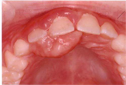
This is an otherwise healthy 8 year-old-boy who underwent a punch biopsy for a small gingival swelling which was removed in March 2007. The lesion recurred in September 2007 in a much larger and more rapidly growing form (Figure 1). This lesion measured 1 X 1.5 cm in size and was red and vascular in color compared to the previously excised lesion, which was 8 X 4 mm in size and pink in color. The current lesion originated between teeth #s 8 & 9. Both lesions were not painful but the recurrent lesion was large enough to interfere with speech and eating. Occlusal radiograph of the first lesion was negative for any bony involvement.
Diagnostic Information Available
This is an otherwise healthy 8 year-old-boy who underwent a punch biopsy for a small gingival swelling which was removed in March 2007. The lesion recurred in September 2007 in a much larger and more rapidly growing form (Figure 1). This lesion measured 1 X 1.5 cm in size and was red and vascular in color compared to the previously excised lesion, which was 8 X 4 mm in size and pink in color. The current lesion originated between teeth #s 8 & 9. Both lesions were not painful but the recurrent lesion was large enough to interfere with speech and eating. Occlusal radiograph of the first lesion was negative for any bony involvement.
Figure 1 This photograph represents the six-month recurrence taken in September 2007. Note the size and color of the palatal gingival swelling. The labial gingiva is also red and slightly swollen.
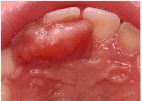
Figure 2 Closer look at the recurrent exophytic red gingival swelling on the palatal gingiva.
The patient is generally in good health and has no significant past medical history.
The patient first presented with an asymptomatic small and pink gingival swelling involving tooth #8 only. It was stated to be of two weeks’ duration. Under local anesthesia, the first lesion was excised in March 2007 at the Department of Oral Medicine, University of Washington. In September 2007, the patient presented with a recurrence of a much larger gingival swelling (Figure 1). The recurrence was much more vascular and thus red in color.
Figure 1 This photograph represents the six-month recurrence taken in September 2007. Note the size and color of the palatal gingival swelling. The labial gingiva is also red and slightly swollen.
Treatment
Under general anesthesia, a full-thickness incision was made around the facial surface of teeth #s 6-9. The lesion was fully excised and the area sutured using 4-0 Vicryl. The surgery left a small denuded area of anterior palate which was packed with iodoform gauze and impregnated with antibiotic ointment. A pre-surgical stent was placed onto the maxillary dentition for pain control and to act as a bandage. The area healed well with no complication or evidence of recurrence.
Incisional and excisional biopsy
The first specimen showed evidence of a moderately cellular fibrous connective tissue mass with an ulcerated surface, clusters of calcified material and areas of osteoid material. The ulcerated surface was covered by fibrin and neutrophils. The mass and the surrounding connective tissue were infiltrated by neutrophils, plasma cells and lymphocytes. The second specimen was more of a loose and vascular granulation tissue mass with small clusters of calcified material and many inflammatory cells.
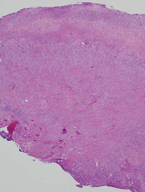
Figure 3 Low power (100X) the H & E stained specimen shows an ulcerated and moderately cellular fibrous connective tissue nodule with clusters of calcified material
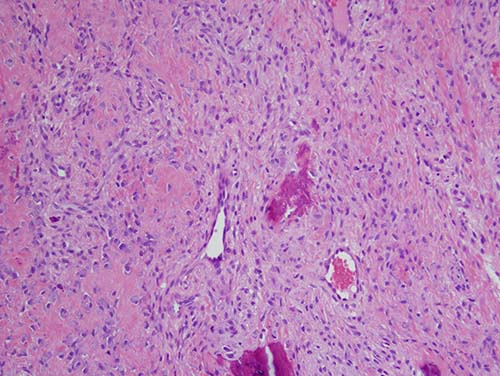
Figure 4 Higher power (200X) the H & E stained specimen shows cellular fibrous connective tissue containing osteoid material, young osteoblasts and clusters of calcified material.
After you have finished reviewing the available diagnostic information