All cases are discussed by: Dr. Dolphine Oda, UW-Oral Pathology Biopsy Service
Large multilocular, expansile radiolucency, anterior mandible
Contributed by:
Drs. Mathew Monaco & John Fink
Oral & Maxillofacial Surgery & Cellnetix Pathology, Palmer, Alaska
Case Summary and Diagnostic Information
This is a 52-year-old female who presented to her dentist with a new onset of left lower jaw swelling associated with left lower lip and chin paresthesia.

This is a 52-year-old female who presented to her dentist with a new onset of left lower jaw swelling associated with left lower lip and chin paresthesia. A clinical exam showed teeth #s 21 and 22 with 1+ mobility and 23 with 2+ mobility. A CBCT was performed and showed a large radiolucency with a hint of multilocular appearance between teeth #s 20-26 (Figure 1). It was well demarcated and was pushing teeth apart. It showed significant buccal expansion with thinning of the cortical bone (Figure 2).
Figure 1 This is a reconstructed panoramic radiograph from CBCT. Note the large radiolucency with well-demarcated margin between teeth #s 20-26. There is a hint of multilocular appearance of this radiolucent lesion.
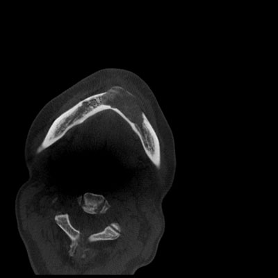
Figure 2 This is an axial view of the CBCT. Note the large and radiolucent lesion in the anterior mandible to left posterior mandible. It is well-demarcated and shows thinning of the cortical bone and significant buccal expansion
The patient’s past medical history is significant for knee surgeries, appendectomy, and hysterectomy.
An incisional biopsy was performed under local anesthesia. The incision was between teeth #s 21 to 24. The area was aspirated with [[return of froth blood]]. The area was unroofed and a portion of the lesion was exposed, curetted, and placed in formalin for pathology reading. The area was irrigated with sterile saline and sutured with 3-0 chromic gut suture.
Histologic examination revealed multiple pieces of soft tissue composed of cellular and vascular granulation tissue containing numerous giant cells and early bone formation (Figure 3). The granulation tissue was cellular and vascular. It contained extravasated erythrocytes and small clusters of hemosiderin pigment. It also contained numerous giant cells which were of variable shapes and sizes and were haphazardly arranged (Figures 4 & 5). New bone formation was identified (Figure 5).
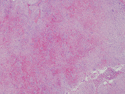
Figure 3 Low power (x40) histology of H & E stained section demonstrating cellular and vascular granulation tissue with many giant cells that are haphazardly arranged.
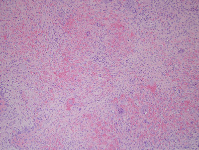
Figure 4 Higher power (x100) histology of H & E stained section shows a closer look at the vascular and cellular granulation. It also shows the giant cells at a closer magnifications.

Figure 5 Higher power (x100) histology of H & E stained section shows a closer look at the vascular and cellular granulation. It also shows the giant cells at a closer magnifications.
After you have finished reviewing the available diagnostic information