Nonhealing ulcer plus red & white lesion of left lateral tongue in nonsmoker
Can you make the correct diagnosis?
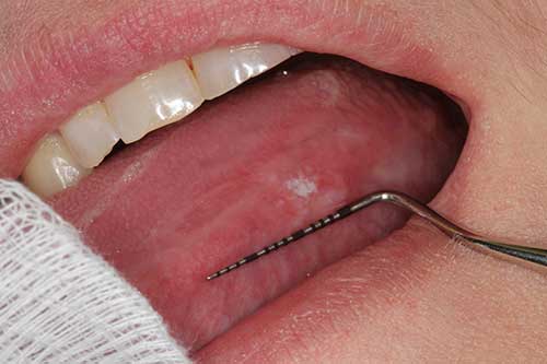
This is a 41-year-old white female who was referred to Dr. Dhaliwal for the evaluation of a red and white lesion with central ulceration on the left border lateral tongue.
Sorry, you are incorrect!
The differential diagnosis of a red and white lesion with or without ulceration on the lateral tongue should include frictional keratosis, i.e., chronic tongue chewing. Frictional keratosis can be of chronic duration and the patient would typically confirm that trauma is present. It can occur at any age and has no gender predilection. It is, however, more common in younger patients. It usually involves the lateral tongue, cheeks, or lips. On clinical examination and palpation, frictional keratosis lesions tend to be white and rough but can also get ulcerated and become red and white if the patient bites the area deeply or forcefully. The patient denied biting her tongue and the histology is not consistent with chronic tongue chewing/biting.
Sorry, you are incorrect!
Although the current case does not have the typical features of traumatic ulcer or its deep variant known as TUGSE, trauma-induced ulceration should be on the differential diagnosis because the site of occurrence is frequently afflicted with surface ulcerations. Traumatic ulcers and TUGSE commonly occur on the lateral (anterior) tongue, usually in patients with a history of trauma i.e. biting their tongue forcefully. Traumatic ulcers are usually painful and heal in two weeks. The duration of one month makes the conventional “traumatic ulcer” unlikely, but not so for TUGSE, since TUGSE can last for eight weeks or more. TUGSE affects patients of all ages but is more common in adult males. The histology is not consistent with traumatic ulcer or the TUGSE variant.
Congratulations, you are correct!
The site of lateral-ventral tongue and the clinical presentation of a red and white lesion with a nonhealing ulcer are typical of the clinical presentation of oral squamous cell carcinoma (SCC) in smokers. However, this patient has no history of tobacco use. The patient’s age and gender are also not consistent with tobacco-associated tongue SCC. The patient’s age suggests oral SCC associated with oncogenic human papilloma virus (HPV), though the site and the gender do not. The immunohistochemistry stain for p16, a surrogate antibody to oncogenic HPV, is negative. The etiology is not definitive at this point, but the patient’s ten-year history of systemic sclerosis is implicated. Systemic sclerosis has been reported, at least in one large cohort study, to lead to a 25-fold higher risk of developing tongue SCC than in the general population. The H & E histology is consistent with SCC.
Sorry, you are incorrect!
It is unusual for oral candidiasis to occur as an isolated lesion on the lateral tongue or in the mouth in general. Among the few exceptions to this rule is median rhomboid glossitis on the dorsal, middle and posterior tongue. It may also occur on the palate under a denture/movable appliance that is worn 24/7 and is not washed properly (inflammatory papillary hyperplasia) and on the bilateral angles of the mouth (angular cheilitis superimposed with candidiasis). Candidiasis isolated to the lateral border of the tongue can occur superimposing an ulcer or eroded epithelium but not as an opportunistic disease. The histology is not consistent with candidiasis.