February 2010: Extensive superficial ulceration of the left ventral tongue
Can you make the correct diagnosis?
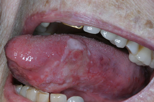
This is an 87-year-old white female referred by her general dentist for the evaluation of a large, irregular, mostly superficial and ulcerated red and white lesion on the left lateral and ventral surfaces of tongue of unknown duration (Figure 1).
Sorry! you are incorrect
The mostly superficial, eroded red-and-white look in an elderly person’s tongue should raise the possibility of a fungal infection such as candidiasis. However, the focal deep ulceration in this case is not consistent with candidiasis; neither is the histology.
Candidiasis is the most common oral opportunistic infection, especially in AIDS patients. It is one of the earliest presentations of immune deficiency, sometimes appearing in otherwise healthy-appearing patients (1-3). If it occurs in non-compromised patients it usually involves the tongue, skin folds, vagina and urinary tract. It may occur in compromised patients, especially in the GI tract, lungs, heart valve, kidneys and meninges. It is caused by Candida albicans, which presents in the form of delicate pseudo hyphae and budding spores; Candida is normally found, in up to 50% of the population, in the oral cavity. It affects high-risk patients such as infants under six months of age for lack of immunity, pregnant females, debilitated patients with chronic diseases, diabetics, patients who are immunosuppressed, and patients on long-term broad-spectrum antibiotics or steroids.
Acute and chronic types are described, including acute pseudomembranous candidiasis, which is found in chronically ill patients and infants. It presents as white, soft, slightly elevated plaques on the buccal mucosa and tongue. It can be wiped out leaving a relatively normal appearing mucosa, or slightly red mucosa. Acute atrophic candidiasis usually presents with thin, atrophic erythematous and painful mucosa. Chronic hyperplastic candidiasis is a firm, white, persistent plaque on the lip, tongue and buccal mucosa. It is difficult to wipe off. Chronic atrophic candidiasis, “denture sore mouth,” angular cheilitis is characterized by diffuse inflammation of denture bearing area. Oral candidiasis usually responds to Nystatin suspension while systemic candidiasis responds to amphotericin B. If the patient does not respond to treatment, test for diabetes or other endocrinopathies.
AIDS patients can develop all types of candidiasis. Atrophic candidiasis can be present all through the course of the HIV infection and is the first to appear. It is usually asymptomatic but with time can be painful. Pseudomembranous candidiasis is common on the palate and the vestibules. It appears during the early stages and it presents as white plaques that can be wiped out. Patients complain of a burning sensation and discomfort in swallowing. The infection may include the pharynx and esophagus. Candidiasis of the latter two types in high-risk individuals is almost pathognomonic for AIDS.
Sorry! you are incorrect
Erosive lichen planus (ELP) or erosive lichenoid drug reaction should be included on the differential diagnosis given the erosive generalized look of this lesion and the age of the patient. However, this is an isolated lesion on the ventral tongue without evidence of any such lesion on the bilateral buccal mucosa and vestibule, making it an unlikely candidate for lichen planus or a lichenoid reaction. The histology in this case is also not supportive of this diagnosis.
Erosive LP is the second most common type of lichen planus (LP) in the mouth (4-5). It is usually symptomatic; patients complain of sensitivity to hot and cold food and beverages, spicy food and alcohol. Clinically, it is hard to distinguish from candidiasis and other mucocutaneous diseases such as mucous membrane pemphigoid and pemphigus vulgaris. The mucosa appears red, thin and ulcerated. At times, radiating white striae are present at the periphery of the lesions.
Lichenoid drug reaction is a common condition, especially in elderly patients. It presents with clinical features similar to erosive lichen planus, mainly on the buccal mucosa (6-7). It is associated with the ingestion of certain medications such as antibiotics, antihypertensive drugs, allopurinol (gout), diuretics, antidiabetics, gold, mercury, antibiotics, and antihistamines. The histology and IMF characteristics of both entities are similar. It is well established that LP of the mouth, especially the erosive type, has a small tendency (up to 2.5%) for transformation in long standing LP that has been present for five years or more. Some LP cases respond to topical steroids, e.g. betamethasone 0.1% cream while others, the more severe type, may require systemic steroid therapy.
Congratulations! You are correct
Oral squamous cell carcinoma of the mouth is a highly aggressive neoplasm that currently ranks as the fifth most common malignant neoplasm worldwide and accounts for an estimated 90% of oral malignancies (8-10). Oral SCC occurs predominantly in males over the age of 40 years, with an observed male-to-female ratio of 2:1 generally and 1.4:1 in the USA (8). Excluding the outer lip, the most common sites (in decreasing order) are the ventral and lateral surfaces of the tongue (25-50%), floor of mouth (15%), gingiva (12%) and palate (9%). The buccal mucosa and retromolar pad areas (3%) have a relatively low incidence of occurrence (9) unless the patient is a chronic smokeless tobacco user. Oral SCC varies in presentation from deceptively innocent looking to obviously malignant. It may present as a non-healing ulcer, or as red, white or mixed red-and-white lesions. Characteristic signs of oral SCC are non-healing ulcer, ulcer with rolled borders, fungation, fixation and induration. Rarely, oral SCC may present as unexplained asymptomatic lateral neck lymphadenopathy (8-10). Oral SCC is most commonly associated with chemically induced mutagenesis, specifically tobacco and alcohol use (9).
Tobacco use is described in over 75% of oral SCC patients (8-10). Tobacco and alcohol have been shown to act synergistically in the development of oral SCC. Human papilloma virus (HPV) has also been found to have a high prevalence in oral cancer, especially in younger patients with no history of tobacco use. Other factors include poor oral hygiene, syphilis, chronic candidiasis, iron and dietary deficiencies, herpes simplex and various other immunologic factors, and lichen planus—especially persistent erosive lichen planus.
Determination of the prognosis of oral SCC is based on its clinical stage and histological classification. Although oral SCC is a diagnosis made by histology, surgeons tend to depend exclusively on the TNM classification system for clinical staging and treatment decisions. Prognosis is dependent on the TNM staging system; the most important prognostic sign is the presence or absence of metastases at the time of diagnosis. The prognosis thus improves when the lesion is detected early. Oral SCC patients die mainly of infection due to lowered resistance or of hemorrhage if the tumor erodes through one of the main blood vessels.
References
- Akpan A, Morgan R. (2002). Oral Candidiasis. Postgraduate Med J, 78:455–459.
- Perezous LF, Flaitz CM, Goldschmidt ME, Engelmeier RL. (2005). Colonization of Candida species in denture wearers with emphasis on HIV infection: a literature review. J Prosthet Dent ; 93: 288–293.
- Webb BC, Thomas CJ, Whittle T. (2005). A 2-year study of Candida-associated denture stomatitis treatment in aged care subjects. Gerontology; 22: 168–176.
- Scully and M. Carrozzo, Oral mucosal disease: lichen planus, (2008). Br J Oral Maxillofac Surg; 46: 15–21
- Eisen D. (2002). The clinical features, malignant potential, and systemic associations of oral lichen planus: a study of 723 patients, J Am Acad Dermatol; 46: 207–214
- Scully C, Bagan JV. (2004). Adverse drug reactions in the orofacial region. Crit Rev Oral Biol Med;15:221–239.
- Neville BW, Damm DD, Allen CM et al. Allergies and immunologic diseases. In: Neville BW, Damm DD, Allen CM, Bouquot JE, eds. Oral & maxillofacial pathology, 3rd edn. New York: W.B. Saunders Company, 2009:347-350.
- Bundgaard T, S Bentzen. (1996). Histopathologic, stereologic, Epidemiologic, and clinical parameters in the prognostic evaluation of squamous cell carcinoma of the oral cavity. Head & Neck; 18:142-152.
- Barasch A, DE Morse. (1994). Smoking, gender, and age as risk factors for site-specific intraoral squamous cell carcinoma. Cancer; 73:509-513.
- Syrjanen SM, KJ Syrjanen. (1988). Human papillomavirus (HPV) DNA sequences in oral precancerous lesions and squamous cell carcinoma demonstrated by in situ hybridization. J Oral Pathol. 17:273.