February 2009: Dark brown to black gingival swelling, right posterior palate
Can you make the correct diagnosis?
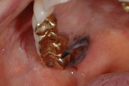
This is an 82-year-old white female whose chief complaint was a painful lesion in the right posterior palate associated with the palatal gingiva of tooth # 2 (Figure 1).
Sorry! you are incorrect
Dark brown to black pigmented lesions of the mouth are usually caused by endogenous (melanin, blood, hemosiderin) or exogenous (metal, carbon) particles. Melanocytic nevi and melanotic macules should be considered given the color of this lesion. However, the rolled border, the lack of uniformity in the coloration and the crater in the middle suggest this lesion is neither a nevus nor an oral melanotic macule. The histology is also not supportive of melanotic macule or any of the melanocytic nevi.
Melanocytic nevi (common moles) are rare in the mouth but are very common on the skin. Most skin nevi are the acquired (developmental) type while congenital nevi are much less frequent on the skin and also far less frequent in the mouth. Nevi are benign and most likely hamartomatous proliferations of cells of neural crest origin which can be melanocytes or other close related cells. In the oral cavity, they most commonly occur on the hard palate, buccal mucosa, gingiva and lips (1-2). They are more common in females around 30 years of age. Intramucosal (intradermal) and blue nevi constitute 80-90% of all oral melanocytic nevi with compound and junctional nevi accounting for less than 10-20% of cases (1-2). Nevi vary in color, from colorless to dark blue, brown or black; some oral nevi lack the brown color. They can be flat, raised, smooth-surfaced or papillary. Histologically, they vary in morphology depending on the type. Nevi are usually not removed unless they are chronically irritated. They are sometimes removed for aesthetic reasons. They should be removed if they change in appearance in accordance with the “A-E rule”: A) for asymmetry, B) for border (irregular border), C) for color (lack of uniformity in color), D) for diameter (larger than six mm in diameter) and E) for enlarging (5). They should be surgically removed if they ulcerate or bleed.
Melanotic macules present as oval-shaped brown and flat lesions less than 8 mm in diameter (3-4), mostly on the lower lip near the midline. Melanotic macules of the lip are called labial melanotic macules and the ones that occur in the oral cavity are called oral melanotic macules. Oral melanotic macules are more common on the gingiva, followed by the buccal mucosa. Oral melanotic macules can occur at any age, with a 2:1 prevalence in females. They usually present as a solitary lesion, but can present in multiples (1-4). When multiple macules are identified, it is necessary to rule out Addison’s disease, Peutz-Jeghers syndrome and other conditions such as drug-induced hyperpigmentation (2-4).
Sorry! you are incorrect
Kaposi’s sarcoma (KS) is more common on the palate and presents as purplish red in color. It can be flat of nodular; all of this would make it reasonable to include it on the differential diagnosis. However, KS in the United States is predominantly associated with AIDS. This patient has no such history. The histology in this case is also not supportive of KS.
Kaposi’s sarcoma, malignant lymphoma, cervical cancer, candidiasis, and deep fungal infections such as histoplasmosis, coccidioidomycosis, and pneumocystis carinii are among the main AIDS-defining malignant neoplasms and infections. KS is the most common of the malignant neoplasms, affecting 50% of AIDS patients and accounting for approximately 90% of all cancers in this group (9-11). However, it must be mentioned that since the introduction of HAART antiviral treatment of AIDS patients, the incidence of KS has gone down (10-11).
About 71% of AIDS-related KS cases affect the oral cavity combined with skin and visceral lesions (9-10). In about 22% of these patients the oral cavity is the primary site for KS and, at times, it is the only affected site. It is a malignant neoplasm of vascular origin believed to be of endothelial origin, mainly lymphatic endothelium. It usually presents in multiple areas all at the same time. Recently, a report concluded that it can be induced by human herpes virus 8 (HHV8) (9-10); in this case, it is obviously oncogenic rather than infectious and reactive in nature. HHV8 has been described in other malignant neoplasms such as AIDS-related body cavity lymphoma.
Four types of KS are described (9-11): classic, AIDS-related, immuno-suppression-associated and the African type. The classic type of KS is a rare and non-aggressive disease and is especially rare in the oral cavity (9). It is usually slow-growing, predominantly affecting older males of Mediterranean origin including Italians, Greeks and Ashkenazi Jews, and occurs as multiple reddish-purple lesions on the lower extremities. AIDS-related KS is aggressive in clinical behavior (9-11) and affects mostly young homosexual and bisexual males in the US and heterosexual males in African countries; it occurs in the oral cavity and on the skin of the head and neck, trunk, etc. The mouth, especially the palate, is a frequent site of occurrence as described above (accounting for 71% of cases). It may present as flat to nodular purplish-red lesion(s). It is usually asymptomatic, but may be painful. The immuno-suppression type is associated with organ transplant patients, especially kidney transplant patients (9-11). It is described in other patients, especially those on low-dose prednisone. It has also been described in cancer patients who are on long-term chemotherapy. This type also affects patients of Mediterranean origin. The African type is endemic in some parts of Africa (9-10), and is further classified into two types: an aggressive and fatal type that occurs in children and a less aggressive form that occurs in adults. The latter is more common on the extremities, while the former is associated with lymphadenopathy and is more generalized. The aggressive form may also be associated with AIDS.
Treatment of KS depends on the clinical subtype and stage, the extent of the disease, whether it presents in singles or multiples, and whether it involves the skin/mucosa or the visceral organs. Many factors are involved in deciding on a specific mode of treatment. Single skin lesions of the classic type respond well to electron beam radiation therapy. Radiation of single oral lesions is not as successful given the oral complications from radiation treatment, including mucositis, xerostomia and osteoradionecrosis (9-11). Local intra-lesional injections have also been described to be useful in treating some single lesions, including those of the oral cavity. Systemic chemotherapy is used for more advanced and disseminated types of the disease. The prognosis varies and depends on the subtype; clearly, classic KS has a much better prognosis than AIDS-related KS or the African type affecting children.
Sorry! you are incorrect
When faced with a dark blue or black pigmented lesion of the mouth, a clinician should consider the possibility that it is the result of exogenous metal or carbon particles, such as an amalgam tattoo. Although amalgam tattoo is common on the gingiva, the rolled border and cratered dark brown presentation suggest that it is not the correct diagnosis in this case. The histology in this case is also not supportive of amalgam tattoo.
Second to racial pigmentation, amalgam tattoo is the most common pigmented lesion in the oral cavity. It is an example of a local deposition of exogenous metallic material from a dental filling, usually the silver metal found in an amalgam (6-8). The fine dark granules of this material are deposited on the very thin collagen fibers of the connective tissue, around small blood vessels, of the basement membrane, between muscle fibers, salivary gland tissue and fat. Amalgam tattoo occurs as a result of metal implantation in lacerated mucosa during a dental filling or the removal of an old amalgam filling (6-8). It can also be the result of amalgam falling in the dental socket following an extraction or in the area of an apicoectomy sealing. Like any cutaneous tattoo, it becomes part of the live tissue and leads to a permanent coloration of the mucosa, usually flat and grey to bluish-black or black in color. Occasionally, it can be exophytic or nodular (6); this is usually the result of a foreign body reaction to the metal. The margins can be irregular, well defined or diffuse, blending in with the surrounding unaffected mucosa. The borders are usually flat and not rolled. They can enlarge and give the impression of lateral spread; this happens during the first months of implantation, especially when a foreign body reaction with macrophages presents, carrying the material laterally and giving the impression of spreading, thus making one think beyond amalgam tattoo to a more sinister disease. Once the area scars, the lateral spread ceases. Amalgam tattoo most commonly occurs on the alveolar mucosa, buccal mucosa, and gingiva (6-7), but has been described on the palate, floor of mouth and tongue. It can be viewed radiographically, especially within a socket if present in clusters. Histologically, it presents as granular black material deposited on the surrounding thin collagen fibrils. Sometimes, foreign body reaction with foreign-body-type giant cells, chronic inflammation and scarring are present. Sometimes, an incisional biopsy is recommended to eliminate the possibility of other, more serious diseases; otherwise, there is no need for treatment.
Congratulations! You are correct
Malignant melanoma (MM), whether on the skin or in the oral cavity, has the same cell of origin: the melanocyte. Despite the common cell origin, however, these lesions have different clinical behavior. Mucosal melanomas are very rare and are more aggressive than those of the skin. They usually affect adults with an average age of 55; occasionally, they affect patients under 30 years of age (12-13). Skin MM used to have a poor prognosis but that has changed significantly; in 1950, the 5-year survival rate for skin MM was 50%, while today it is 90% (12-14). However, mucosal melanomas still carry a dismal 5-year survival rate of not higher than 38% (12). Skin MM, like mucosal melanoma, occurs mostly in adults around 50 years of age and rarely affects children.
Cutaneous malignant melanoma is the third most common skin epithelial malignancy (following basal cell carcinoma and squamous cell carcinoma) but by far the most aggressive of the three. In the United States alone, over 50,000 cases are diagnosed annually with about 8,000 dying of the disease each year (12). It has been on the increase since 1960 and has recently plateaued. In the 1960s, 1 in 600 individuals were expected to develop MM in their lifetime, compared with 1 in 71 in 2001. Australia has the highest number of skin MMs, a number that has been doubling every 10 years (14). The etiology of MM and its rate of increase are complex and are beyond the scope of this discussion. However, it is important to mention that multiple factors seem to play a role the development of the MM and its progression. The most important factor is exposure to ultraviolet light, which explains cases on the skin but does not explain the oral, nasal, and other non-sun exposed areas that develop MM. It has been suggested that the age of the individual at the time of exposure is important (12-14): it is also suggested that sunburns, especially blistering sunburns in childhood, increase the risk of developing MM in adulthood. Genetics play a significant role in MM, especially in patients born in families with a history of this disease. For instance, patients from families with a history of dysplastic mole syndrome or patients who have a first relative with malignant melanoma are at a higher risk of developing the disease (12-14). 70% of cutaneous MMs occur in a preexisting pigmented nevus. Pigmented nevi are common and have a one in a million chance of transformation to MM. This does not necessitate surgical removal, but rather suggests that practitioners should be attentive to alterations in nevi such as changes in color, shape and size; ulceration and/or bleeding; inflammation; pain; and a diameter change of greater than 7mm. These are the clinical criteria used in England (5) to assess the clinical presentation of MM. Bleeding and ulceration of a pigmented nevus should be viewed seriously, and the lesion should be soon biopsied or completely removed and submitted for microscopic evaluation. The clinical features of MM in the United States follow the ABCDE rule: asymmetry, borders, color, diameter and enlargement. A pigmented lesion that is asymmetrical with irregular borders (especially borders with notching) and uneven coloration (a range of tan to dark brown as is the case with our patient), is larger than 6 mm in diameter, and continues to enlarge should be viewed with suspicion and biopsied immediately.
The oral mucosa is rarely affected by MM and for that reason, the etiology of oral malignant melanoma is still unclear. Local melanosis and tobacco use have been implicated. Race also appears to play a role; for example, 7.5% of all reported oral melanomas affect those of Japanese descent and 10% affect those of Ugandan descent, compared with less than 1% of cases occurring in Caucasians. Cutaneous MM, however, occurs at a much higher rate in Caucasians than in those with brown or black skin, with a rate of 1:83 in whites compared to 1:1176 in dark-skinned individuals (13). Mucosal melanomas are extremely rare compared to cutaneous melanomas; they constitute less than 1% of all MMs. For that reason, it is always prudent to first rule out metastatic MM to the oral cavity from the skin when a biopsy of an oral pigmented lesion confirms the diagnosis of MM. 80% of oral MMs affect the palate and the maxillary gingiva. Mandibular gingiva can be affected, but rarely. Cutaneous MMs occur mostly on the interscapular area of the back and back of the legs (12). 25% occur in the head and neck area, including the face, eyes, nose and mouth (14). In the oral cavity, they present as flat, multiple, graded or with uneven coloration, and irregular borders; they are sometimes ulcerated. They are usually asymptomatic; pain is described only rarely.
Histologically, MMs demonstrate radial and vertical growth patterns. The vertical pattern is important because it plays an important role in predicting prognosis; for that reason, the depth of invasion is always reported. Depth of invasion in skin MMs is fully established following Clark and Breslow’s staging according to depth (12). Obviously, the deeper the tumor invasion, the higher the microscopic staging and the worse the clinical behavior. Determining prognosis is not simple, though; other factors play a role in worsening the prognosis, such as the location, age and gender of the patient. Surgery is the treatment of choice treatment for MM of the oral cavity. Radiation and chemotherapy may be used in advanced cases.
References
-
- Meleti M, Vescovi P, Mooi WJ, van der Waal I. Pigmented lesions of the oral mucosa and perioral tissues: a flow-chart for the diagnosis and some recommendations for the management. Oral Surg Oral Med Oral Pathol Oral Radiol. 2008 May;105:606-16.
- Buchner, P.W. Merrel and W.M. Carpenter, Relative frequency of solitary melanocytic lesions of the oral mucosa, J Oral Pathol Med 33 (2004), pp. 550–557
- Eisen, Disorders of pigmentation in the oral cavity, Clin Dermatol 18 (2000), pp. 579–587.
- Çiçek and Ü. Ertas¨, The normal and pathological pigmentation of oral mucous membrane: a review, J Contemp Dent Pract 3 (2003), pp. 76–86
- Healsmith, MF, Bourke, JF et al. An evaluation of the revised seven-point check list for the early diagnosis of cutaneous malignant melanoma. Br J Dermatol 1994; 130: 48-50.
- Buchner A. Amalgam tattoo (amalgam pigmentation) of the oral mucosa: clinical manifestations, diagnosis and treatment. Refuat Hapeh Vehashinayim. 2004 Jul;21(3):25-8.
- Owens BM, Johnson WW, Schuman NJ. Oral amalgam pigmentations (tattoos): a retrospective study. Quintessence Int. 1992 Dec;23(12):805-10.
- Shah, G, Alster, TS. Treatment of an amalgam tattoo with a Q-switched alexandrite (755 nm) laser. Dermatol Surg 2002; 28: 1180-1181.
- Buchner Brown EE, Fallin D, Ruczinski I, Hutchinson A et al. Associations of classic Kaposi sarcoma with common variants in genes that modulate host immunity. Cancer Epidemiol Biomarkers Prev. 2006 May;15(5):926-34.
- Lager I, Altini M, Coleman H, Ali H. Oral Kaposi’s sarcoma: a clinicopathologic study from South Africa. Oral Surg Oral Med Oral Pathol Oral Radiol Endod. 2003 Dec;96(6):701-10.
- Fletcher CDM. Diagnostic histopathology of tumors. Vascular tumors. Third Edition. Churchill-Livingston, 2008. Pages 58-62.
- Hicks MJ, Flaitz CM. Oral mucosal melanoma: epidemiology and pathobiology. Oral Oncol. 2000 Mar; 36(2):152-169.
- Rapidis, AD, Apostolidis, C et al. Primary malignant melanoma of the oral mucosa. J Oral Maxillofac Surg. 2003; 61: 1132-1139.
- Rigel, DS, Friedman, RJ et al. The incidence of malignant Melanoma in the United States: issue as we approach the 21st century. Am J Acad Dermatol 1996; 34: 839-847.