Large Multilocular, Expansile Radiolucency, Right Posterior Mandible
Dolphine Oda, BDS, MSc
doda@u.washington.edu
Dr. Francis Collins
Wenatchee Oral & Maxillofacial Surgery, Wenatchee, WA
Case Summary and Diagnostic Information
This is a 26-year-old Hispanic female who presented to her dentist with mild pain and very large swelling on the right posterior mandible.
Diagnostic Information Available
This is a 26-year-old Hispanic female who presented to her dentist with mild pain and very large swelling on the right posterior mandible. The area was described to be of a problem for six months, but the swelling increased tremendously within the last a few weeks. The clinical findings are those of a large submandibular facial swelling. Intra-orally (Figure 1), the swelling expands buccal and lingually pushing teeth #s 30 and 31 buccally, more so tooth #30 with extreme mobility of the two teeth. The panoramic view shows a large and expansile multilocular radiolucency extending from the apex of tooth #27 to inferior border of mandible and to the right mandibular notch. Inferior border of mandible is obscured from the multilocular appearance (Figure 2).
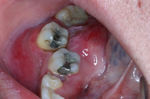
Figure 1. This photograph was taken at first clinical presentation. Note the buccal and lingual expansion and the redness of the mucosa and gingiva overlying the swelling. Teeth #s 30 & 31, especially tooth #31 are pushed buccally.
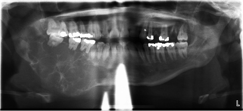
Figure 2. This radiograph was taken at first clinical presentation. Note the ballooning expansion and the multilocular radiolucency extending from tooth #27 anteriorly to the mandibular notch posteriorly. Also, note the resorption of teeth #s 30-32.
The patient’s past medical history is unremarkable.
The patient reported severe swelling and loosing of teeth #s 30 and 31 over the last several weeks. The area however has not been feeling well and has been progressively enlarging over the last six months.
Treatment
Under local anesthesia, an incisional biopsy was performed. After the incisional biopsy, the patient was referred for surgical excision of the neoplasm.
Excisional Biopsy
Histologic examination revealed multiple fragments of soft tissue composed epithelial islands of variable shapes and sizes lined by one layer of palisaded columnar cells with reversed polarization (Figures 3 & 4). The islands are filled with stellate reticulum-like epithelial cells in some areas and squamous epithelium in others (Figures 3 & 4).
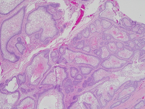
Figure 3. Low power (x100) H & E histology shows epithelial islands of variable shapes and sizes lined by one layer of palisaded columnar epithelial cells with reversed polarization. The center of the islands contains stellate-reticulum-like epithelial cells in some areas and squamous in others.
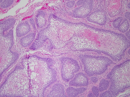
Figure 4. Higher power (x200) H & E histology shows epithelial islands with one layer of palisaded columnar epithelial cells and center filled with stellate-reticulum-like epithelial cells in some areas and squamous epithelium in others.
After you have finished reviewing the available diagnostic information