Return to Case of the Month Archives
Large radiolucency left maxilla and left maxillary sinus
Dolphine Oda, BDS, MSc
doda@u.washington.edu
Contributed by
Dr. Gerald Dolgash
Oral Surgery Associates, Olympia, Washington
Case Summary and Diagnostic Information
This is a 12-year-old Hispanic girl who was referred by her general dentist for a large swelling in the left maxilla. It involved teeth #s 11-15 and the maxillary sinus (Figure 1). The lesion was of unknown duration and was 2 x 4 cm in size. The soft tissue overlying the lesion was reddish in color and the swelling was both buccal and palatal. Teeth #s 12-14 were mobile. The CT scan showed the tumor to have infiltrated the roots of the left maxillary permanent dentition. Her past medical history is unremarkable.
Diagnostic Information Available
After you have finished reviewing the available diagnostic information
This is a 12-year-old Hispanic girl who was referred by her general dentist for a large swelling in the left maxilla. It involved teeth #s 11-15 and the maxillary sinus (Figure 1). The lesion was of unknown duration and was 2 x 4 cm in size. The soft tissue overlying the lesion was reddish in color and the swelling was both buccal and palatal. Teeth #s 12-14 were mobile. The CT scan showed the tumor to have infiltrated the roots of the left maxillary permanent dentition. Her past medical history is unremarkable.
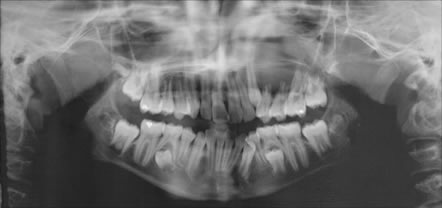
Figure 1 Panoramic view at first presentation demonstrating large unilocular radiolucency of the left posterior maxilla, displacing and resorbing teeth and extending into the maxillary.
Her past medical history is unremarkable.
Panoramic radiograph demonstrated a large and destructive radiolucency of the left posterior maxilla. Teeth #s 12 and 13 show evidence of resorption. The lesion involves the maxillary sinus and is displacing teeth (Figure 1). Clinical examination revealed a large swelling expanding the buccal and palatal bone in the area; it also revealed the mobility of teeth #s 12-14.
Figure 1 Panoramic view at first presentation demonstrating large unilocular radiolucency of the left posterior maxilla, displacing and resorbing teeth and extending into the maxillary.
Treatment
Under IV sedation, the area was biopsied. The lesion was described as solid and hemorrhagic. The specimen was submitted for histologic evaluation.
Incisional and excisional biopsy
Histological examination revealed multiple large pieces of vascular granulation tissue with many giant cells (Figures 2 & 3). The latter were of variable shapes and sizes; some were closely associated with small blood vessels while most were haphazardly arranged. Small clusters of extravasated erythrocytes and hemosiderin pigment were identified (Figure 3).
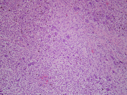
Figure 2 Low power (x100) histology shows a mass of vascular granulation tissue with many multinucleated giant cells of variable shapes and sizes, clusters of hemosiderin pigment and some extravasated erythrocytes.
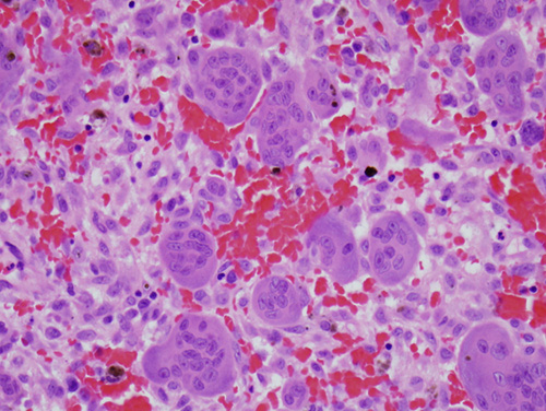
Figure 3 Higher power (x200) demonstrating many giant cells, granulation tissue background, hemosiderin pigment and extravasated erythrocyte.