All cases are discussed by: Dr. Dolphine Oda, UW-Oral Pathology Biopsy Service
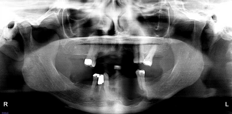
Large multilocular, expansile radiolucency, anterior mandible
Contributed by: Dr. Benjamin Johnson
Oral & Maxillofacial Surgery, Auburn, WA
Case Summary and Diagnostic Information
This is a 46-year-old female who presented to her dentist with loose anterior teeth and a large swelling in the area of tooth #22 involving the anterior mandible and crossing the midline to area of tooth #27 (Figure 1).
This is a 46-year-old female who presented to her dentist with loose anterior teeth and a large swelling in the area of tooth #22 involving the anterior mandible and crossing the midline to area of tooth #27 (Figure 1). This lesion is of unknown duration and was described to be 3x3x3 cm in size. Clinically, the lesion presented as a large swelling with a red/purple and brown appearance.
Figure 1 This radiograph was taken at first clinical presentation. Note the multilocular radiolucency with well-demarcated margin between teeth #s 22 and 27.
The patient’s past medical history is significant for sleep apnea and prediabetes.
The patient reported loose teeth in the anterior mandible around tooth #22. In that area, there was a large swelling that crossed the midline in the anterior mandible to involve tooth #27. The lesion was well-demarcated radiolucency, multilocular radiographically, with clearly defined margins. It was clinically described as a swelling with “inferior border intact.” This is consistent with the radiographic findings in which the inferior border of the mandible is mostly intact.
Under local anesthesia, an incisional biopsy was performed. After the incisional biopsy, the patient was referred for surgical excision of the neoplasm.
Histologic examination revealed multiple fragments of soft tissue composed of a neoplasm of odontogenic origin. It is made up of epithelial islands of variable shapes and sizes lined by one layer of palisaded columnar cells with focal reversed polarization (Figures 2 & 3). The islands are filled with stellate reticulum-like epithelial cells in some areas and squamous epithelium in others (Figure 4).
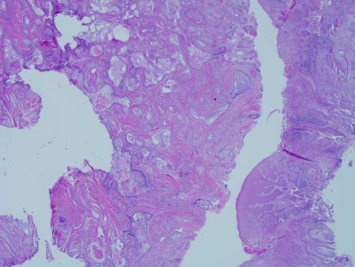
Figure 2 Low power (x40) H & E histology shows epithelial islands of variable shapes and sizes suspended on mature connective tissue background. The center of the islands contains stellate-reticulum-like epithelial cells in most areas and squamous cells focally.
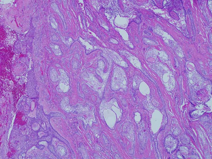
Figure 3 Higher power (x100) H & E histology shows epithelial islands of variable shapes and sizes suspended on mature connective tissue background. The center of the islands contains stellate-reticulum-like epithelial cells in most areas and squamous cells focally.
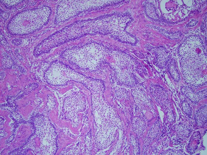
Figure 4 High power (x200) H & E histology shows epithelial islands with one layer of palisaded columnar epithelial cells with focal reversed polarization. The center of the islands is predominantly filled with stellate-reticulum-like epithelial cells with focal areas of squamous epithelium.
After you have finished reviewing the available diagnostic information