December 2010: Single Dome-Shaped Swelling on the Upper Lip
Can you make the correct diagnosis?
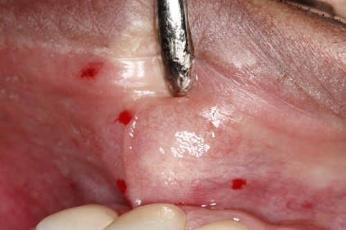
This is a 33-year-old male who presented with a smooth-surfaced, pink, firm and movable nodule on the upper lip, left side (Figure 1). It is of two years’ duration. It is around two cm in diameter at its greatest dimension and is not painful. The swelling is otherwise asymptomatic with an intact mucosa.
Sorry! you are incorrect
A movable, firm nodule of the upper lip should make a clinician think of canalicular adenoma since the upper lip is the most common location for that neoplasm. The age and gender in this case is not typical, but it should nonetheless be on the differential diagnosis. The histology is not supportive of canalicular adenoma.
Canalicular adenoma is a benign neoplasm that is almost exclusively of minor salivary gland origin. The upper lip is the most common location, accounting for over 80% of all cases (1-3). Occurrences in the parotid gland and other major salivary glands are described, but rarely. They present as well-circumscribed, firm, movable, slow-growing, painless nodules (1-3). They occur in females over the age of 50 and are usually single nodules. However, canalicular adenomas are sometimes known to present in a multifocal manner, which a clinician may mistake for invasion or metastasis (1). They are treated by simple enucleation (1-3). Histologically, canalicular adenomas present as an encapsulated neoplasm with cords, duct-like structures of monotonous, cuboidal or low columnar basaloid cells, and sometimes with large cystic dilatation of the duct-like spaces (1-3).
Congratulations! You are correct
Although the age, gender and location in this case are not supportive of pleomorphic adenoma, the histology was typical of it.
Pleomorphic adenoma is the most common benign salivary gland neoplasm of both the major and minor salivary glands. It originates from the myoepithelial cells and the reserve cells of the intercalated ducts. It accounts for 80% of all benign salivary gland neoplasms. It occurs in both major and minor salivary glands and accounts for up to 77% of parotid, 68% of submandibular, and 43% of minor salivary gland tumors (4-6). It is most common in females 30-50 years of age, but it is also described in children (4). One study reports 1% of cases affecting children under 10 years of age and 5.9% between the ages of 10-20 (4, 6). It presents as a small, painless, slowly enlarging nodule. If left untreated, it can enlarge significantly, sometimes increasing by several pounds in weight (3-4). It occurs in the oral cavity, especially the palate and lips (4-6). On the palate, it is usually located in the posterior hard palate or anterior soft palate but can also be in the posterior soft palate; PA usually occurs in the posterior and lateral palate, as opposed to torus palatines, which usually occurs in the middle hard palate and in the anterior. The posterior hard palate mixed tumor is fixed due to the bone-bound anatomy of the region; the tumor is otherwise movable. Histologically, mixed tumor has a wide variety of cellular and pattern manifestations; the main cellular components are epithelial duct-like structures and mesenchymal-like tissue such as myxochondroid matrix. These lesions are generally encapsulated, ranging from predominantly myxoid (36%) to extremely cellular (12%) (4-6). Complete surgical removal with clean margins is the preferred treatment (4-6). Palatal lesions respond well to excision in one piece with the periosteum and overlying mucosa. Pleomorphic adenoma has a good prognosis, but it has a tendency for recurrence (up to 44%) if not treated thoroughly (3-4). The risk of recurrence is less if it occurs in the minor salivary glands (up to 20%). The risk of malignant transformation is about 5% (3-6).
References
- Mansueto G, Falleti J, De Cecio R, Papa F, De Rosa G. Synchronous bilateral multifocal canalicular adenoma: a case report of an unusual finding. Clin Exp Dermatol. 2009 Dec;34(8):e587-9. Epub 2009 May 22.
- Azevedo LR, Dos Santos JN, De Lima AA, Machado MA, Grégio AM. Canalicular adenoma presenting as an asymptomatic swelling of the upper lip: a case report. J Contemp Dent Pract. 2008 Jan 1;9(1):91-7.
- Pons Vicente O, Almendros Marqués N, Berini Aytés L, Gay Escoda C. Minor salivary gland tumors: A clinicopathological study of 18 cases. Med Oral Patol Oral Cir Bucal. 2008 Sep 1;13(9):E582-8.
- Toida M, Shimokawa K, Makita H, Kato K, Kobayashi A, Kusunoki Y, Hatakeyama D, Fujitsuka H, Yamashita T, Shibata T. Intraoral minor salivary gland tumors: a clinicopathological study of 82 cases. Int J Oral Maxillofac Surg. 2005 Jul;34(5):528-32. Epub 2005 Jan 24.
- Yih WY, Kratochvil FJ, Stewart JC. Intraoral minor salivary gland neoplasms: review of 213 cases. J Oral Maxillofac Surg. 2005 Jun;63(6):805-10.
- Hamakawa H, Takarada M, Ito C, Tanioka H. Bone-forming pleomorphic adenoma of the upper lip: report of a case. J Oral Maxillofac Surg. 1997 Dec;55(12):1471-5.
- Hashiba Y, Nozaki S, Yoshizawa K, Noguchi N, Nakagawa K, Yamamoto E. Recurrent multinodular neurilemmoma of the female upper lip. Int J Oral Maxillofac Surg. 2007 Feb;36(2):171-3.
- Yang SW, Lin CY. Schwannoma of the upper lip: case report and literature review. Am J Otolaryngol. 2003 Sep-Oct;24(5):351-4.
- Anastassov GE, van Damme PA. Angioleiomyoma of the upper lip: report of a case. Int J Oral Maxillofac Surg. 1995 Aug;24(4):301-2.
- Maeda Y, Hirota J, Osaki T, Hayashi K, Sonobe H, Otsuki Y. Angiomyoma of the upper lip: report of a case with electron microscopic and immunohistochemical observation. Br J Oral Maxillofac Surg. 1989 Jun;27(3):236-42.
Sorry! you are incorrect
A movable, firm nodule of the upper lip should make a clinician think of schwannoma since the lips are a relatively common location for this neoplasm. The age in this case is typical of this neoplasm. The histology is not supportive of schwannoma.
Schwannoma is a benign, firm, smooth-surfaced, encapsulated and mobile neoplasm of Schwann cell origin. It occurs at any age but is more common in individuals 30-50 years of age with equal sex distribution (7-8). The tongue is the most common location, followed by the floor of mouth and the lips (7-8). It is also described as occurring within the jaw bones, especially the mandible. Up to 48% of schwannomas occur in the head-and-neck area. They are usually isolated lesions unless they present as part of neurofibromatosis type 1. Shwannoma presents as a slow-growing, firm, rubbery, smooth-surfaced nodule (7-8). The histology is usually diagnostic where the lesion is encapsulated and has two patterns: Antoni A, the cross section of which gives rise to “Verocay bodies,” and Antoni B, which is loose and resembles neurofibroma. This lesion is encapsulated while neurofibromas are usually not encapsulated. Simple excision is the treatment of choice and recurrence is rare.
Sorry! you are incorrect
A movable, firm nodule of the upper lip should make a clinician think of leiomyoma or angiomyoma (vascular leiomyoma). The age and gender in this case is typical of leiomyoma. The histology, however, is not supportive of leiomyoma.
Leiomyoma is a benign neoplasm of smooth muscle origin. Uncommon in the oral cavity, it mainly occurs in the G.I. tract, uterus and skin. In the oral cavity, it most likely originates from the vascular smooth muscle. Vascular leiomyoma, also known as angiomyoma, accounts for 75% of oral leiomyomas. Patients tend to be over 30 years of age and are predominantly males. It is most common on the posterior tongue, palate, cheeks and lips. It is a slow growing, painless, pedunculated, smooth-surfaced, normal color or slightly bluish nodule. Intra-osseous leiomyomas have been reported, but very rarely. Histopathologically, this neoplasm is made up of a well-circumscribed to encapsulated nodule composed of bundles of smooth muscle cells, some surrounding blood vessels in cases of angiomyoma. Simple and conservative excision is the treatment of choice.