August 2016: Gingival swelling, lingual gingiva between teeth #s 24 & 25s
Can you make the correct diagnosis?
Case Summary and Diagnostic Information
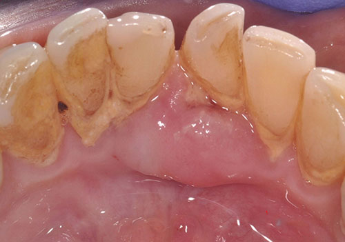
This is a 64-year-old male who presented with a painful, mostly smooth-surfaced pink gingival swelling.
Sorry, you are incorrect!
The exophytic, pink, sessile and firm nature of this lesion are all consistent with those of a benign reactive gingival swelling responding to local stimuli such as excessive plaque or calculus, as is apparent in this patient’s mouth. Reactive gingival swellings include pyogenic granuloma, inflammatory fibrous hyperplasia, peripheral ossifying fibroma and peripheral giant cell granuloma.
Although pyogenic granuloma (PG) is the most common reactive gingival swelling, it should not be considered in this case because of the color of the lesion. Pyogenic granulomas tend to be vascular and red in color. For the same reason, peripheral giant cell granuloma (PGCG) should not be considered. They usually present as red, gray, or purplish in color.
Inflammatory fibrous hyperplasia (IFH) is a likely reactive gingival swelling to consider. It is on the differential diagnosis because this lesion clinically presents at any age and gender. Inflammatory fibrous hyperplasia is common and is usually exophytic, firm, sessile, smooth surfaced, and pink like the surrounding gingiva if it is not ulcerated. It occurs more commonly on the gingiva but can occur elsewhere in the mouth.
Peripheral ossifying fibroma (POF) is another type of reactive gingival swelling that should be under consideration. It tends to present on the anterior gingiva in form of a sessile firm nodule, often ulcerated, but sometimes smooth surfaced and pink as is the case with this patient. POFs are more common on the buccal gingiva but can occur on the lingual (palatal) gingiva or between teeth. They are more common in young females, occurring in females at a ratio of 3:2 with a typical age range of 10-20 years. Although the clinical presentation and site of this case are typical of POF, the age and gender are not consistent with this condition.
The histology in this case is not consistent with IFH, POF, PG or PGCG.
Sorry, you are incorrect!
Exophytic, sessile, pink and firm gingival swellings may also be benign neoplasms of tooth (odontogenic) origin that solely occur on the gingiva; however, such neoplasms are rare. The clinical presentation of an exophytic, sessile, pink, and smooth-surfaced swelling on the gingiva in this case is consistent with the clinical presentation of peripheral ameloblastoma, as is the patient’s age and gender. The site in the anterior gingiva is unusual since peripheral ameloblastomas tend to occur more often on the posterior mandibular gingiva.
Ameloblastoma is one of the most common benign neoplasms of odontogenic origin. It accounts for 11% of all odontogenic neoplasms. Almost 99% of ameloblastomas are intra-osseous (central/within bone) and about 1% are present completely outside the bone, usually in the gingiva; these are called peripheral ameloblastomas. Peripheral ameloblastomas are a slow-growing and non-aggressive counterpart of intra-osseous ameloblastomas. They present as small, well-circumscribed, firm nodules on the gingiva, mainly of the posterior mandible in patients around 50 years of age. They are slightly more common in males. They are usually asymptomatic, pink like the surrounding mucosa, smooth surfaced, and sessile.
The histology in this case is not consistent with peripheral ameloblastoma or any other peripheral odontogenic neoplasm.
Sorry, you are incorrect!
Generally, benign soft tissue neoplasms such as solitary neurofibromas and leiomyomas are rare in the mouth. If they do occur, the gingiva is not a common site. On the rare occasions that leiomyomas occur in the mouth, they tend to be of the vascular type (angiomyoma). Vascular leiomyomas are slightly reddish-gray in color and they, too, rarely occur on the gingiva.
Solitary neurofibromas of the oral cavity can occur between 20 and 30 years of age. Some studies find they are slightly more common in females, while others report an equal gender predilection. They usually occur on the buccal mucosa, tongue, palate and, rarely, on the gingiva. They are non-tender, smooth surfaced, slow-growing swellings that may be soft or firm.
Leiomyomas of the oral cavity tend to originate from the smooth muscle cells around blood vessels. These are known as vascular leiomyomas (or angiomyomas) and account for 75% of oral leiomyomas. Patients tend to be over 30 years of age and are predominantly males. They more commonly occur on the posterior tongue, palate, cheeks and lips. They are slow growing, painless, sessile or pedunculated, smooth surfaced, and normal colored or slightly bluish nodules.
The histology in this case is not consistent with peripheral ameloblastoma or any other peripheral odontogenic neoplasm.
Congratulations, you are correct!
Although the clinical presentation is that of a benign and most likely reactive swelling, given the patient’s history of leiomyosarcoma of the extremities and later of the kidney and lover, one has to include metastatic disease on the differential diagnosis. Cancer metastasis to the oral cavity is neither specific nor common; such cases constitute less than 1% of all oral malignant neoplasms. They can be devastating to the patient because metastasis to other sites has already developed or is inevitable. Theoretically, any malignant neoplasm can metastasize to the oral cavity, but in actuality few do and of the ones that do, the majority is carcinomas rather than sarcomas. The most common malignant neoplasms that metastasize to the mouth are from the breast, lung, prostate and kidney. Malignant neoplasms from the colon, pancreas, esophagus, thyroid, cervix, and liver have also been described. Breast cancer is the most common neoplasm to metastasize to the oral cavity altogether. Lung and prostate cancers are the most common neoplasms to metastasize to the oral cavity in men. In most cases, the oral presentation is a secondary diagnosis when the primary diagnosis of malignancy in a distant organ has been already made and the patient has had or is undergoing treatment for it. In rare cases, the oral lesion is the first manifestation of the disease. By far the most common location is the posterior mandible, where 80% of cases occur, followed by the gingiva. This condition is mostly described in adults over the age of 30 and rarely in children.
There are very few cases described in the literature of leiomysarcoma metastasizing to the oral cavity. Most of these cases occurred in women with known Stage IV leiomyosarcoma of the uterus.
This case would represent an extremely rare minority of leiomyosarcoma cases metastasizing from the extremities to the mouth in a male patient.
The histology in this case is consistent with metastatic leiomyosarcoma in both years 2014 and 2016 specimens.