All cases are discussed by: Dr. Dolphine Oda, UW-Oral Pathology Biopsy Service
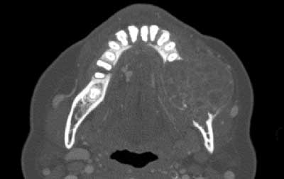
Large multilocular, expansile radiolucency left posterior mandible
Contributed by: Drs. Namou Kim & Lee-Ching Zhu
Swedish Hospital-ENT and Kaiser Permanente-Pathology, Seattle, WA
Case Summary and Diagnostic Information
This is a 54-year-old female who presented with a chief complaint of an enlarging left mandible.
This is a 54-year-old female who presented with a chief complaint of an enlarging left mandible. The swelling is of 2-3 months duration. The patient reports having significant pain, mobility of teeth and difficulty chewing. She has stopped chewing food relying on a puréed and liquid diet at this time. The patient also feels that her dental occlusion has shifted but denies any paresthesia of the left lower lip. The CT scan of the neck shows a very large multilocular mass (Figure 1) occupying the entire left body of the mandible. This lesion has essentially eroding both lingual and buccal cortical bones.
Figure 1 This CT image was taken by the ENT surgeon prior to surgery. The ballooning multilocular radiolucent expansion is impressive. Note the thinning and perforation of the buccal and lingual cortical bone.
The patient’s past medical history is significant for hypertension, dysfunctional uterine bleeding and hyperlipidemia.
This patient presented with an enlarging and painful swelling of the left mandible (Figure 1) that was of 2-3 months duration. The patient stopped chewing food secondary to the pain and tooth mobility and relied on a puréed and liquid diet for the duration of the swelling. The CT images show that the lesion has essentially eroded both lingual and buccal cortical bone of the left mandible.
An incisional biopsy was taken from the mandible. The pathology repot suggested that brown tumor of hyperparathyroidism be ruled out. She had a calcium level drawn, which was high normal at 9.5. The parathyroid hormone level was 19 making it less likely to be brown tumor secondary to hyperparathyroidism. The swelling showed significant bone loss and the tooth mobility was described to have features of “teeth floating in space.” The lesion was completely resected (Figure 2) and the area reconstructed with a fibular free flap. The cut section of the gross specimen was solid and cystic with spaces filled with erythrocytes (Figure 3).
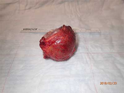
Figure 2 This photograph depicts gross appearance of the surgical specimen of the swelling identified in figure 1. It is small and red in color. It represents a conservative resection of the swelling along with two molar teeth.
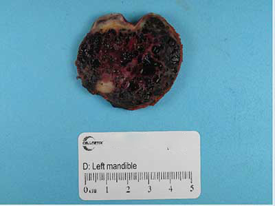
Figure 3 This photograph represents the surgical specimen cut in half with cut surface demonstrating a lesion with combined solid and cystic areas. The cystic areas are filled with blood as noted by the dark brown color of the cut surface.
Histologic examination revealed multiple fragments of soft tissue composed of highly dilated vascular spaces surrounded by cellular granulation tissue (Figure 4) with many giant cells and focal areas of a benign fibro-osseous type reaction. Some of the vascular spaces were filled with erythrocytes (Figures 4 & 5). The granulation tissue surrounding the spaces was cellular and contained giant cells of variable shapes and sizes (Figure 6). In focal areas, the histology was that of a fibro-osseous type reaction. This tissue was composed of small foci of calcified material surrounded by cellular fibrous connective tissue (Figure 7).

Figure 4 Low power (x100) H & E histology illustrates a highly vascular lesion made up of large spaces, some filled with erythrocytes. These spaces are surrounded by cellular granulation tissue.
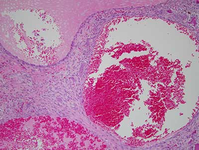
Figure 5 Higher power (x200) H & E histology illustrates a large sinusoidal space, some containing erythrocytes surrounded by cellular granulation tissue with multinucleated giant cells.
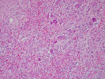
Figure 6 Higher power (x 200) H & E histology illustrates cellular fibrous and granulation tissue with giant cells and calcified material.
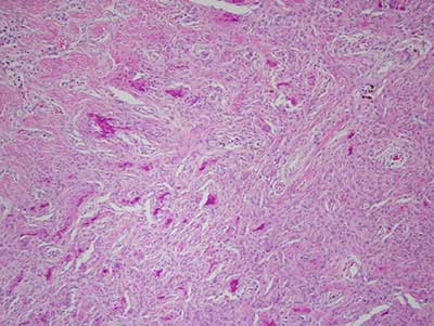
Figure 7 Higher power (x 200) H & E histology illustrates the part of the lesion which was more fibro-osseous in nature. Note the cellular connective tissue and calcified material.
After you have finished reviewing the available diagnostic information