Mixed radiolucent radiopaque lesion associated with impacted tooth #32
Can you make the correct diagnosis?
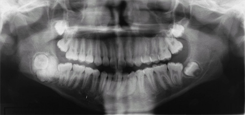
This is a healthy 13-year old African American male who first presented for an initial orthodontic examination. Clinical examination of the area showed a normal range of the mandibular motion with no evidence of bony expansion. The overlying and surrounding oral mucosa was unremarkable.
Congratulations, you are correct!
Odontoma, whether complex or compound, hamartoma or neoplasm, is the most common odontogenic tumor, accounting for 22% of them (1-4). It is of mixed epithelial and mesenchymal origins. It is usually associated with unerupted teeth (1-4). It can occur at any age, but is most common in the first two decades of life, with an average age of 14 or 18. It is slightly more common in females and more common in the maxilla, especially the anterior maxilla, than in the mandible (4). Compound odontomas are more common in the anterior jaws, while complex odontomas in the posterior jaws (2, 4). In about 80% of cases, they are associated with impacted or unerupted teeth. Radiographically, odontomas present as a well-circumscribed radiolucency resembling a dental follicle or dentigerous cyst with one or multiple radiopaque pieces resembling teeth (1). Compound odontomas tend to occur between teeth and tend to be composed of multiple small tooth-like structures, while complex odontomas tend to occur in the posterior jaws and present as a conglomerate mass. Both types are made up of enamel matrix, dentin, cementum, and dental pulp surrounded by a dental follicle or cyst. Histologically, the tooth-like structures are arranged in a uniform manner similar to the normal tooth. The structures in complex odontomas are mixed and disorganized. These lesions are benign and are conservatively treated with simple curettage (1-4). Recurrence is not described; if it recurs, one must rule out other odontogenic lesions such as calcifying odontogenic cyst, ameloblastic fibro-odontoma and others.
Treatment
In anticipation of an increased risk of jaw fracture, the patient had orthodontic brackets and a passive arch wire with lugs placed so that following surgery he could be placed in intermaxillary fixation if indicated. Under general anesthesia the patient was prepared and draped for an intraoral surgical procedure. A mucoperiosteal flap was elevated from the necks of the bicuspid and molar teeth in the right mandible, extending superiorly up the ramus to expose the lateral aspect of the mandible. A wedge of soft tissue was excised posterior to the second molar and submitted as part of the specimen. The bone overlying the lesion was removed and was distinctly seen as a calcified mass, which was subsequently sectioned and removed in multiple pieces, separating easily from the adjacent bone. The sectioning revealed fragments of what appeared to be small malformed teeth, although the mass was generally amorphous.
The third molar was also distinctly involved with the mass. Complete removal of the mass left a smooth walled bony defect (Fig 2). The inferior alveolar nerve was intact and displaced inferiorly following the complete removal of the mass. The large defect was packed with a proprietary iodoform dressing and the mucoperiosteal flap repositioned and closed with resorbable sutures.
The patient was placed in intermaxillary fixation at the completion of the surgery. It was noted that despite the dramatic radiographic appearance, there was more bone remaining than anticipated.

Figure 2. This is a panoramic view of the area immediately after surgery. Note the large radiolucent cavity in the area of the impacted tooth and associated tumor.
References
- Vengal M, Arora H, Ghosh S. Large erupting complex odontoma: a case report. J Can Dent Assoc. 2007 Mar;73(2):169-73.
- Noonan VL, Gallagher G, Kabani S. Compound and complex odontomas. J Mass Dent Soc. 2006 Fall;55(3):40.
- Ogunlewe MO, Adeyemo WL, Ladeinde AL. Surgical management of a large complex odontoma of the mandibular angle-ramus region through intra-oral buccal approach–A case report. Niger Postgrad Med J. 2005 Dec;12(4):312-5.
- Chen Y, Li TJ, Gao Y. Ameloblastic fibroma and related lesions: a clinicopathologic study with reference to their nature and interrelationship. J Oral Pathol Med. 2005 Nov;34(10):588-95.
- Dhanuthai K, Kongin K. Ameloblastic fibro-odontoma: a case report. J Clin Pediatr Dent. 2004 Fall;29(1):75-7.
- Oghli AA, Scuto I, Ziegler C. A large ameloblastic fibro-odontoma of the right mandible. Med Oral Patol Oral Cir Bucal. 2007 Jan 1;12(1):E34-7.
- Gallana-Alvarez S, Mayorga-Jimenez F, Torres-Gomez FJ. Calcifying odontogenic cyst associated with complex odontoma: case report and review of the literature. Med Oral Patol Oral Cir Bucal. 2005 May-Jul;10(3):243-7.
- Praetorius F, Hjorting-Hansen E, Gorlin RJ, Vickers RA. Calcifying odontogenic cyst: range, variations and neoplastic potential. Acta Odonotol Scand 1981;39:227-240.
- Gorlin RJ, Pindborg JJ, Clausen FP, Vickers RA. The calcifying odontogenic cyst: a possible analogue of the cutaneous calcifying epithelioma of Malherbe. Oral Surg Oral Med Oral Pathol 1962;15:1235-1243.
- Hong SP, Ellis GL, Hartman KS. Calcifying odontogenic cyst: a review of ninety-nine cases with reevaluation of their nature as cysts or neoplasms, the nature of ghost cells, and subclassification. Oral Surg Oral Med Oral Pathol 1991;72:56-64.
Sorry. You are Incorrect!
The clinical and radiographic findings are consistent with a calcifying odontogenic cyst especially those that are associated with odontomas (7). The histology, however, is not supportive of this diagnosis. Calcifying odontogenic cyst (COC) constitutes around 1% of all odontogenic cysts (7-9). Praetorius et al (8) were the first to classify COC into a simple cyst and a solid neoplastic spectrum. They classified the cystic type into subtypes A, B and C: A denoting a simple cyst, B an odontoma-producing cyst, and C a cyst with ameloblastomatous proliferating. COC tends to occur around the third decade, with a patient age range of 7-82 years (9-10). It occurs equally in the maxilla and mandible, usually anterior to the first permanent molar, though it has a predilection for occurrence in the maxilla in the younger age range. It occurs equally in males and females (9-10). COC occurs more commonly in bone, but it can also occur in soft tissue (in the gingiva without a bony component, also known as peripheral COC (8-10)). The peripheral counterpart of both lesions is less aggressive than the intra-osseous type (9). Histologically, COC ranges from simple cyst with ghost cells and focal ameloblastoma-like epithelial changes to more proliferative epithelium with ghost cells, to calcifications (both amorphous, tooth-like and calcified ghost cells) and to a true odontoma associated with such cyst. Radiographically, COCs tend to be well-defined radiolucencies with occasional small radiopacities. They can be present at the apex, between teeth, or associated with impacted teeth (8-10). Clinically, both COC can expand the jaws and extend into the surrounding soft tissues and even the maxillary sinus. They can also resorb and displace teeth. Treatment of choice is thorough curettage.
Sorry. You are Incorrect!
In clinical presentation, the radiographic changes are consistent with an ameloblastic fibro-odontoma. The histology, however, is not supportive of this diagnosis.
The ameloblastic fibroma is believed to be described by Kruse in 1891. It is a relatively uncommon neoplasm. It is a benign, slow-growing, expansile, neoplasm or hamartoma of mixed epithelial and mesenchymal odontogenic origins (4-6). Some suggest that this lesion may represent an early stage of an odontoma. However, it behaves like a neoplasm in terms of growth and expansion of the jaw bones. It is known to inhibit tooth eruption and is therefore often associated with impacted teeth. It can also displace teeth. Therefore, these lesions can go undetected by parents unless a noticeable expansion occurs or because of failure of tooth eruption and displacement of teeth. It is rarely painful. It occurs most commonly in children in the first and second decade of life with an age range of infancy to 42 years of age (6). It has equal gender predilection and is more common in the posterior mandible than in the maxilla. It also occurs more often in the posterior jaws than in the anterior jaws. Radiographic findings include an impacted/unerupted tooth with a well-defined, usually unilocular radiolucency around the crown of a tooth, similar to a dentigerous cyst. It may also present as a radiolucency containing radiopaque material ranging from flecks to small tooth-like structures. Because of the continued slow growth, this lesion can reach very large sizes if left untreated. The histology of ameloblastic fibro-odontoma is made up of mixed epithelial and mesenchymal components (5-6). The epithelial component presents in the form of small islands, cords and rosettes of epithelial cells with columnar and palisaded peripheries and with centers that may or may not contain stellate or cuboidal epithelial cells. The mesenchymal component is made up of primitive connective tissue stroma with tooth-like hard material that has features of a complex odontoma. They are benign and well-differentiated neoplasms; for that reason, transformation is rarely described. Conservative curettage and enucleation is the treatment of choice, and there is a very low recurrence rate (4-6). The involved tooth is usually extracted. It has a good prognosis.