Return to Case of the Month Archives
Expansile and Radiolucent Lesion in the Left Anterior Maxilla
Dolphine Oda, BDS, MSc
doda@u.washington.edu
Contributed by
Dr. Ed Truelove, Dr. Thomas Morton, Ms. Negin Bardideh
Department Oral Medicine, University of Washington
Case Summary and Diagnostic Information
This is a 28-year-old white male who presented with a bony bump above teeth #s 10 and 11. This lesion was first noticed by his general dentist in September 2004 and later by the oral surgeon on 10/30/2005. It was expansile, especially labially. The bone supporting the two teeth was compromised, but the teeth were not mobile. The area was biopsied on 11/02/2005. The area was described as a solid mass with a gelatinous consistency. It was 2 x 1.5 cm in size. The lesion did not perforate into the maxillary sinus or the nasal cavity. The roots of teeth #s 10 and 11 were devoid of bone on the interproximal surfaces and a significant amount of the labial cortex was missing. The patient is on follow-up in 3-month intervals.
Diagnostic Information Available
This is a 28-year-old white male who presented with a bony bump above teeth #s 10 and 11. This lesion was first noticed by his general dentist in September 2004 and later by the oral surgeon on 10/30/2005. It was expansile, especially labially. The bone supporting the two teeth was compromised, but the teeth were not mobile. The area was biopsied on 11/02/2005. The area was described as a solid mass with a gelatinous consistency. It was 2 x 1.5 cm in size. The lesion did not perforate into the maxillary sinus or the nasal cavity. The roots of teeth #s 10 and 11 were devoid of bone on the interproximal surfaces and a significant amount of the labial cortex was missing. The patient is on follow-up in 3-month intervals.
The patient has type 2 diabetes controlled with medications, including daily insulin injections. The patient is otherwise in good general health.
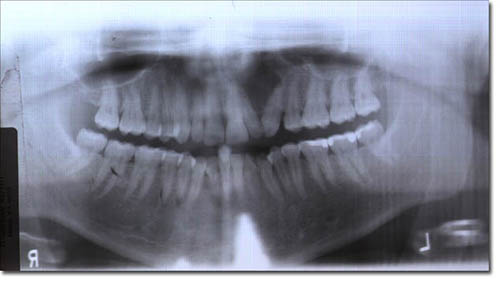
The primary panoramic radiograph (Fig 1) demonstrated a well-demarcated radiolucency between teeth #s 10 and 11. This lesion caused divergence of the two teeth and also expanded the labial bone. Both teeth are vital and show no evidence of caries. The postoperative panoramic view (Fig 2) was taken on March 15, 2006, almost four and a half months after the surgery. It shows healing of the area with a small radiolucency at the apices of teeth #s 10 and 11. The teeth are less divergent.
Figure 1. This is a panoramic radiograph taken at presentation in October 2005. It shows a radiolucency between teeth #s 10 and 11. Teeth #s 10 and 11 are clearly divergent.
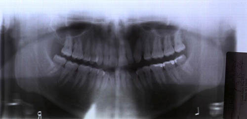
Figure 2. This is a panoramic radiograph taken in March 2006, almost four and a half months after surgery. Note the healing and the less divergent teeth.
Histologic examination revealed multiple pieces of myxoid connective tissue devoid of a lining epithelium (Fig 3). The connective tissue is made up of spindle-shaped and stellate fibroblasts intermixed with short collagen bundles (Fig 4) in most parts and suspended on a myxoid background. Small blood vessels were scattered throughout the specimen.
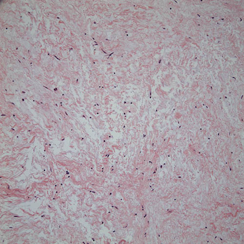
Figure 3. Low power (x100) histology shows a mass of myxoid background with low cellularity, myxoid background intermixed with short collagen bundles consistent with odontogenic myxoma (myxofibroma).
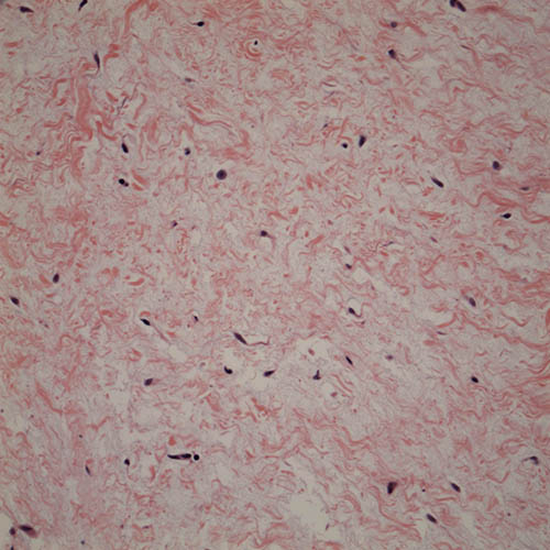
Figure 4. High power (x400) histology shows myxoid background, low cellularity and abundant short collagen bundles.
After you have finished reviewing the available diagnostic information