Return to Case of the Month Archives
Single grayish blue swelling associated with an implant
Dolphine Oda, BDS, MSc
doda@u.washington.edu
Case Summary and Diagnostic Information
This 66-year-old white male presented to the Department of Periodontology, School of Dentistry complaining of a swelling of one year’s duration associated with an implant in the area of tooth #28.
Diagnostic Information Available
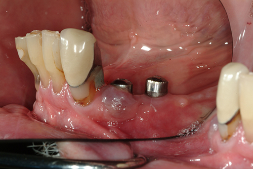
This 66-year-old white male presented to the Department of Periodontology, School of Dentistry complaining of a swelling of one year’s duration associated with an implant in the area of tooth #28 (Figure 1). The patient wears a mandibular partial denture and has Branemark implants in the area of teeth #s 28 and 29 which were placed in the summer of 2003. The patient complained of an unstable mandibular partial denture causing frequent soft tissue ulceration. Excluding the gingival swelling, the soft tissue ulcers healed when the partial denture was left out. This swelling was not painful but the size and color were of concern to the patient. Also of note was that the lesion easily bled with irritation, especially while brushing.
Figure 1. At first visit the gingival swelling around implant of tooth # 28 area was clearly apparent. Note the swelling to be exophytic, sessile, purplish-gray and mostly on the buccal aspect of the implant.
The patient’s past medical history is significant for a heart attack in January 2006. He is currently on Vytorin (ezetimibe/simvastatin, 10mg daily), Altace (Ramipril, 10mg daily), aspirin (80mg daily), Hydrochlorothiazide (12.5mg daily) and daily multivitamins. His past medical history is also significant for cigarette smoking, though he quit in 1977.
At presentation, a grayish-purple soft and asymptomatic swelling of one year’s duration was identified to be associated with a Branemark implant in the area of tooth #28 (Figure 1). At probing, the lesion easily bled, which was also reported to be the case with tooth brushing. Probing around the implant measured 3 mm on the lingual , 5 mm on the mesial, 7 mm on the buccal and 5 mm on the distal.
Figure 1. At first visit the gingival swelling around implant of tooth # 28 area was clearly apparent. Note the swelling to be exophytic, sessile, purplish-gray and mostly on the buccal aspect of the implant.
Histologic examination revealed a piece of soft tissue covered by ulcerated surface epithelium and supported by a mass of granulation tissue containing many giant cells (Figure 3). The latter were of variable shapes and sizes and were haphazardly arranged. Small clusters of hemosiderin pigment were present in the superficial lamina propria (Figure 4). The surface epithelium showed a large ulceration covered by fibrin and neutrophils.
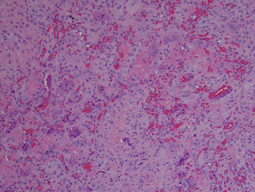
Figure 3. Low power (x100) histology shows a mass of vascular granulation tissue with many multinucleated giant cells.
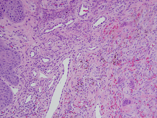
Figure 4. Higher power (x200) histology shows small clusters of hemosiderin pigment in the superficial lamina propria.
After you have finished reviewing the available diagnostic information