Return to Case of the Month Archives
Large, Necrotic Exophytic Mass, Left Posterior Mandible
Dolphine Oda, BDS, MSc
doda@u.washington.edu
Contributed by
Drs. Todd Carter & Ross Beirne
University of Washington, Seattle, Washington
Case Summary and Diagnostic Information
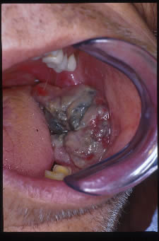
The patient is a 62-year-old white male who presented to the University of Washington emergency room complaining of mouth pain and limited ability to swallow his medications. He also complained of shortness of breath which decreased when he sat down. A large mass was noted to be growing out of the socket of an extracted lower left molar tooth
Diagnostic Information Available
The patient is a 62-year-old white male who presented to the University of Washington emergency room complaining of mouth pain and limited ability to swallow his medications. He also complained of shortness of breath which decreased when he sat down. A large mass was noted to be growing out of the socket of an extracted lower left molar tooth (Fig 1). The tooth had been extracted recently in Alaska. The patient reported increased pain in the area and that the molar tooth had been “abscessed” for approximately one year prior to the extraction. He has a history of smoking one to two packs of cigarettes per day for forty years. He does not consume alcohol.
Figure 1. The left posterior mandibular alveolar ridge demonstrates a large, exophytic purplish-red mass extending downward into the floor of mouth growing in an area of non-healing tooth socket.
The patient is from Alaska and was diagnosed with a Pancoast tumor three months prior to his presentation at the University of Washington. He was in Seattle to visit family and presented at the emergency room with jaw pain. He reported that he had lost 30 pounds over the past year. A chest x-ray showed a 7 X 5 X 6cm posterior apical mass on the right. He had reportedly completed a course of carboplatin and Taxol therapy combined with radiation therapy. He also reported having difficulty swallowing, and choking on liquids. His past medical history also included hypertension, hyperlipidemia, and chronic back pain secondary to an industrial accident and two spinal surgeries.
The patient was a thin, elderly man with a disheveled appearance. He was noted to have Horner’s syndrome on the right side of his face. An oral examination found multiple missing teeth and an upper complete denture (Fig 2). He had full mandibular range of motion. The left posterior alveolar ridge showed a large, ulcerated, necrotic and exophytic mass extending downward into the floor of mouth. The lesion was foul-smelling. A panoramic radiograph (Fig 2) demonstrated irregular bony destruction leading to the saucerization of the alveolar ridge. However, further irregular bone destruction is noted below this concavity, extending downward. In addition to the bony destruction, a large exophytic soft tissue mass is identified radiographically. Reactive bone is noted medial to the lesion and on both sides of the mandible where there appears to be a defect from the wisdom teeth.
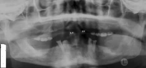
Figure 2. Panoramic view demonstrating upper complete denture and mandibular multiple missing teeth. Left posterior mandible shows evidence of bone destruction and an exophytic soft tissue mass.
The specimen shows multiple pieces of friable soft tissue with sheets of neoplastic cells and fragments of necrotic material. The neoplastic cells show evidence of cellular and nuclear pleomorphism (Fig 3). Some of the cells are large with multiple nuclei while others are small and poorly differentiated (Fig 4). The morphology of the cells is consistent with an epithelial malignancy not native to the oral cavity and for that reason a diagnosis of metastatic carcinoma was rendered. The histology is also consistent with original specimen of poorly differentiated carcinoma of lung.

Figure 3. Low power (x100) histology shows sheets of malignant cells of epithelial origin. They demonstrate cellular and nuclear pleomorphism. The histology is consistent with poorly differentiated malignant neoplasm of epithelial origin.
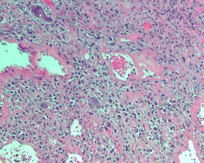
Figure 4. Low power (x100) histology shows sheets of malignant epithelial cells, focal areas of necrosis and malignant cells with multiple nuclei. The histology is consistent with poorly differentiated lung cancer metastasizing to the oral cavity.
After you have finished reviewing the available diagnostic information