Return to Case of the Month Archives
Widespread rough white lesions of the mouth
Dolphine Oda, BDS, MSc
doda@u.washington.edu
Contributed by
Dr. Douglas Wilson, Yakima Oral Surgery
Case Summary and Diagnostic Information
This is a 20-year old white male who presented to his dentist with a 1-year history of widespread white lesions involving several oral sites including the bilateral buccal mucosa, bilateral vestibules and upper and lower lips.
Diagnostic Information Available
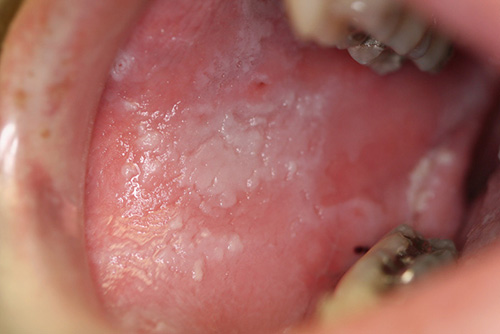
This is a 20-year old white male who presented to his dentist with a 1-year history of widespread white lesions involving several oral sites including the bilateral buccal mucosa, bilateral vestibules and upper and lower lips. At the time of presentation, the lesions (Fig 1& 2) were diffuse, corrugated to verrucoid and rough in consistency. There were areas of focal ulceration associated with a mild burning sensation.
Figure 1. This clinical photograph was taken at presentation. Note the diffuse white corrugated lesions of the right buccal mucosa.
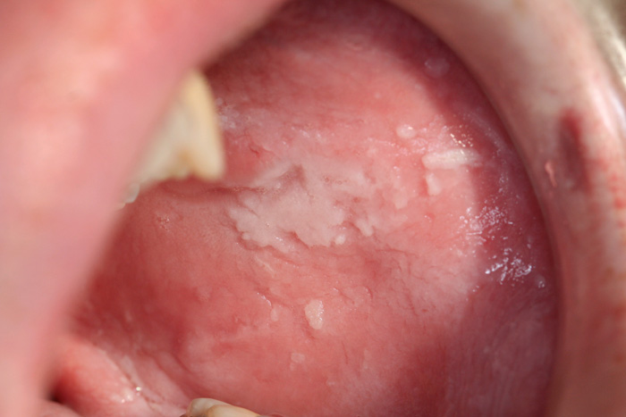
Figure 2. This clinical photograph was taken at presentation. Note the diffuse white corrugated lesions of the left buccal mucosa.
His past medical history is negative for AIDS, diabetes, and alcohol use, but it is positive for cigarette smoking, which he began at age 17, smoking 4-5 cigarettes per day for almost three years. He quit smoking as these lesions began to appear, which was 1 year prior to the incisional biopsy performed by an oral surgeon in September 2006. His family history is positive for similar lesions in his mother’s mouth and in a cousin from the mother’s side of the family.
The oral mucosa was diffusely white and rough (Fig 1 & 2). It involved bilateral buccal mucosa, bilateral vestibules and upper and lower lips. The lesions were rough to palpation and were corrugated to verrucoid. They were focally and mildly symptomatic, described by the patient as a mild burning sensation. The burning sensation corresponded to areas of focal ulceration.
Figure 1. This clinical photograph was taken at presentation. Note the diffuse white corrugated lesions of the right buccal mucosa.
Figure 2. This clinical photograph was taken at presentation. Note the diffuse white corrugated lesions of the left buccal mucosa.
Histologic examination revealed an elongated piece of oral mucosa covered by keratinized epithelium and supported by fibrous connective tissue (Fig 3). The epithelium was of increased thickness and was covered by a thick layer of parakeratin. The spinous layer, especially the upper layers, showed epithelial cells with evidence of cytoplasmic clearing with perinuclear keratin condensations (Fig 4).
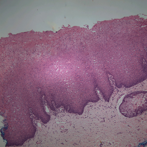
Figure 3.Low power (x100) histology shows a fragment of oral mucosa with thick spinous layer covered by a thick layer of parakeratin. Note the epithelial cells throughout the spinous layer especially in the superficial layers exhibiting keratin condensations around the nuclei.
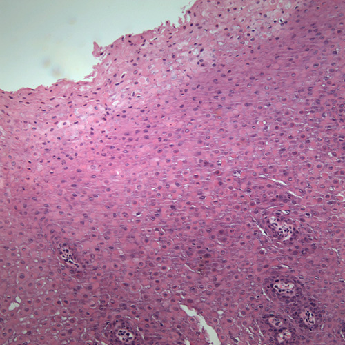
Figure 4. Low power (x100) histology shows a fragment of oral mucosa with thick spinous layer covered by a thick layer of parakeratin. Note the epithelial cells throughout the spinous layer especially in the superficial layers exhibiting keratin condensations around the nuclei.
After you have finished reviewing the available diagnostic information