Return to Case of the Month Archives
Large swelling of the floor of mouth in a newborn baby
Dolphine Oda, BDS, MSc
doda@u.washington.edu
Contributed by
Drs. Pardeep Brar and Mark Egbert
University of Washington and Seattle Children’s Hospital
Case Summary and Diagnostic Information
This is an 11-day-old baby who was presented by her parents to the Oral & Maxillofacial Surgery clinic at Children’s Hospital and Medical Center. Their chief complaint was that the baby’s tongue was progressively increasing in size, making it difficult for the mother to continue breastfeeding.
Diagnostic Information Available
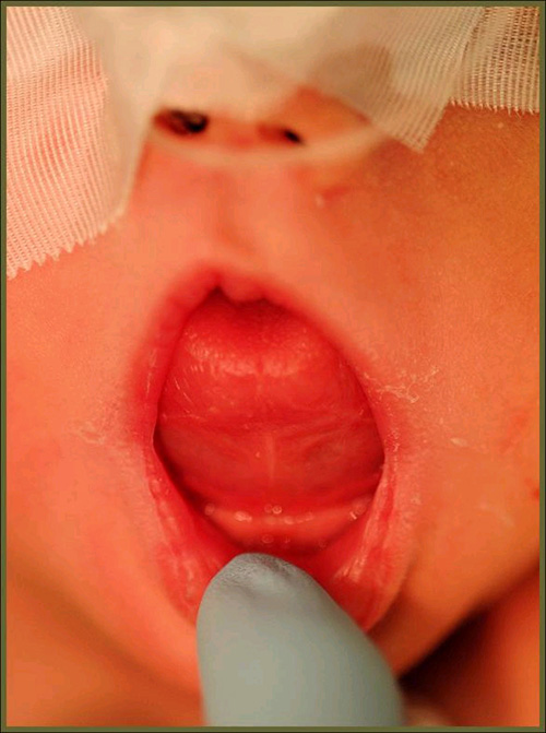
This is an 11-day-old baby who was presented by her parents to the Oral & Maxillofacial Surgery clinic at Children’s Hospital and Medical Center. Their chief complaint was that the baby’s tongue was progressively increasing in size, making it difficult for the mother to continue breastfeeding. The baby did not appear to be in any acute distress. On examination, the tongue was elevated (Fig 1) and there was swelling in the submental space. Palpation of the area revealed a firm mass in the floor of the mouth and posterior tongue. The overlying mucosa was intact and there was no evidence of ulceration or purulent drainage.
Figure 1. This is a clinical photograph as the patient first presented to the Children’s Hospital Dental Clinic. Note the smooth tongue being pushed upward and outward.
This patient’s past medical history is not significant and there are no known drug allergies or developmental defects. She is not on any medications.
The patient presented with an enlarging mass in the tongue. Her tongue was elevated at the primary clinical presentation (Fig 1). There was swelling in the submental space. On palpation, a mobile and firm mass was present in the midline floor of mouth. The color was pink, similar to the surrounding mucosa. There was no ulceration or purulent drainage. The patient was not cyanotic and was able to breath through her nose.
Figure 1. This is a clinical photograph as the patient first presented to the Children’s Hospital Dental Clinic. Note the smooth tongue being pushed upward and outward.
The histology was diagnosed by Dr. Raj Kapur, Seattle Children’s Hospital, Department of Pathology. Histologic evaluation revealed a large lesion of vascular origin (Fig 3). It was made up of vascular spaces of variable shapes and sizes. They are lined by one layer of flat endothelial cells. Some of the spaces contained lymph material with a few lymphocytes while other had clusters of neutrophils (Fig 4) along with some plasma cells.
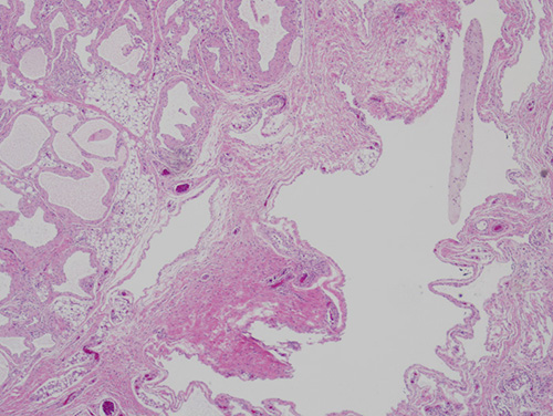
Figure 3. Low power (x100) histology shows thin vascular spaces made up of lymphatic spaces lined by one layer of flat endothelial cells.
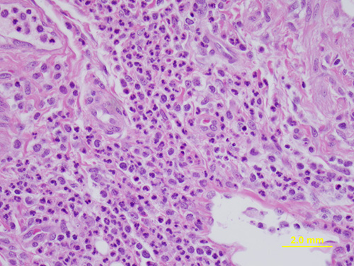
Figure 4. High power (x200) histology shows lymphatic spaces and connective tissue stroma infiltrated by inflammatory cells. They were more in the connective tissue stroma. The inflammatory cells were predominantly neutrophils.
After you have finished reviewing the available diagnostic information