Return to Case of the Month Archives
Single smooth surfaced and well-demarcated nodule, ventral tongue
Dolphine Oda, BDS, MSc
doda@u.washington.edu
Contributed by
Drs. Shradha Bansal, Ryan Gibson & Mark Egbert
Tacoma Dental & Seattle Children’s Hospital, WA
Case Summary and Diagnostic Information
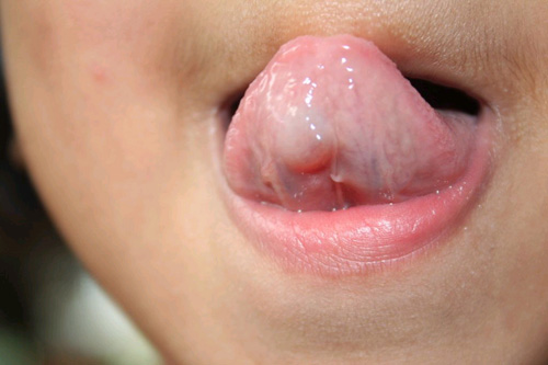
This is a healthy four-year-old girl whose mother was the first to notice a single, smooth-surfaced, well-demarcated and grayish-pink nodule on the right side of the ventral surface of tongue about 4 X 4 mm in size.
Diagnostic Information Available
This is a healthy four-year-old girl whose mother was the first to notice a single, smooth-surfaced, well-demarcated and grayish-pink nodule on the right side of the ventral surface of tongue about 4 X 4 mm in size. It was asymptomatic and enlarged progressively over a period of six to eight weeks to about 1 cm in its greatest dimensions. At early presentation, the mass was described as superficial and fluctuant, although it eventually became firmer. The overlying mucosa was stretched and smooth with no evidence of ulceration.
Figure 1. This is a clinical photograph as the patient first presented to the Children’s Hospital Dental Clinic. Note the dome-shaped smooth surfaced exophytic grayish nodule on the ventral tongue.
Her past medical history is not significant for any diseases or allergies.
This is a healthy four-year-old girl whose mother was the first to notice a single, smooth-surfaced, well-demarcated and grayish-pink nodule on the right side of the ventral surface of tongue about 4 X 4 mm in size. It was asymptomatic and enlarged progressively over a period of six to eight weeks to about 1 cm in its greatest dimensions. At early presentation, the mass was described as superficial and fluctuant, although it eventually became firmer. The overlying mucosa was stretched and smooth with no evidence of ulceration.
The histology was diagnosed by Dr. Laura Finn, Seattle Children’s Hospital; Department of Pathology. The histologic evaluation revealed a cyst-like structure lined by granulation tissue (Fig 3) and filled with mucoid material and foamy macrophages (Fig 4). This structure was focally lined by skeletal muscle bundles and minor salivary gland tissue exhibiting focal fibrosis and chronic inflammation.
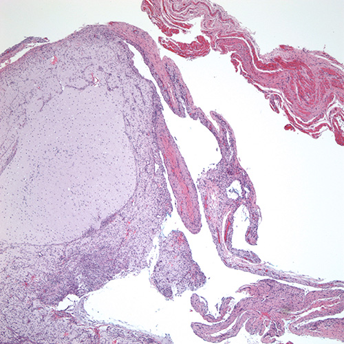
Figure 3. Low power (x100) histology shows a cyst-like structure lined by granulation tissue and filled with mucoid material and foamy macrophages. The lesion was focally surrounded by skeletal muscle bundles.
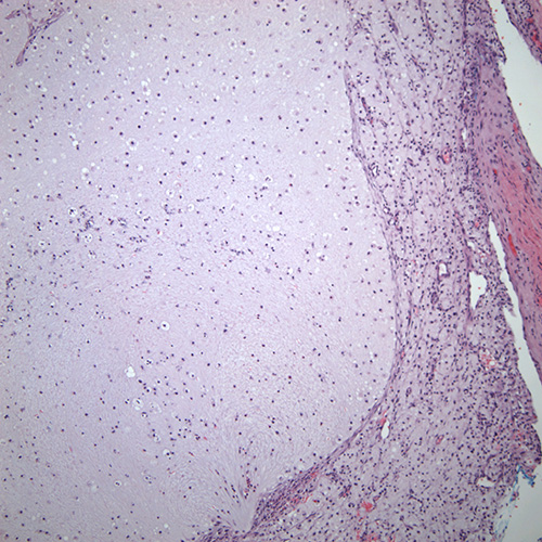
Figure 4. High power (x200) histology shows closer look of the cyst-like structure with the granulation tissue rim and lumen filled with mucoid material and foamy macrophages.
After you have finished reviewing the available diagnostic information