Return to Case of the Month Archives
Gingival swelling of the lingual anterior aspect of the mandible.
Dolphine Oda, BDS, MSc
doda@u.washington.edu
Contributed by
Dr. Walter Kegel, Seattle-Periodontics
Seattle, Washington
Case Summary and Diagnostic Information

This is a 23-year-old pregnant Vietnamese female who presented with this lesion that had been present for two months at the time of her examination.
Diagnostic Information Available
This is a 23-year-old Vietnamese female who, at the time that she presented with this lesion, stated that it had been present for two months (Figures 1 & 2). She was in her seventh month of pregnancy at the time. The lesion did not resolve upon giving birth. This was her first pregnancy, and there had been no significant medical findings or complications. Extensive calculus accumulation was noted throughout the mouth, but especially on the lingual aspect of the mandibular anterior teeth where the lesion is. The lesion was completely excised along with the gross calculus approximately 2 months after delivery. Extensive bleeding was noted upon removal.
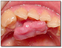
Figure 1. Photograph is taken at first clinical presentation while the patient is seven month pregnant. The lesion is at the anterior lingual mandibular gingiva. It is a large, exophytic, lobular and red mass.
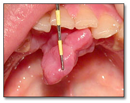
Figure 2. Photograph is taken at first clinical presentation. The lesion is pedunculated as demonstrated by the periodontal probe.
The past medical and family history is non-contributory.
The patient was a young Vietnamese female in her seventh month of pregnancy with extensive and general dental calculus, especially in the anterior lingual mandible. She also had generalized gingivitis. On oral examination a large, exophytic, lobular, red and pedunculated mass of about 2 cm in diameter was found in the anterior lingual mandible (Figures 1 & 2). The lesion was of two months’ duration and was asymptomatic. It was vascular and firm at palpation and was not ulcerated. The lesion was on the lingual gingiva only and did not involve the bone.
Figure 1. Photograph is taken at first clinical presentation while the patient is seven month pregnant. The lesion is at the anterior lingual mandibular gingiva. It is a large, exophytic, lobular and red mass.
Figure 2. Photograph is taken at first clinical presentation. The lesion is pedunculated as demonstrated by the periodontal probe.
The excisional biopsy revealed multiple pieces of soft tissue made up of a mass of vascular granulation tissue covered by intact stratified squamous epithelium (Figure 3). The granulation tissue contained many congested and dilated blood vessels surrounded by proliferative endothelial cells and young fibroblasts (Figures 4 & 5).
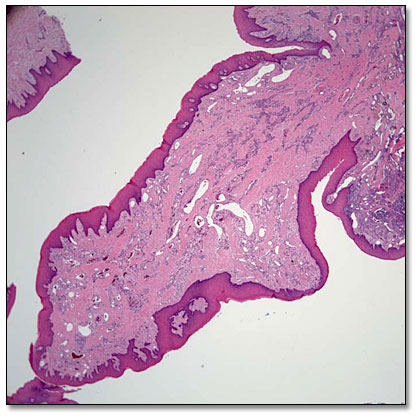
Figure 3. Scanning power (x20) histology shows an exophytic mass of vascular granulation tissue covered by intact stratified squamous epithelium.

Figure 4. Low power (x100) histology shows a mass of vascular granulation tissue consistent with a pyogenic granuloma.
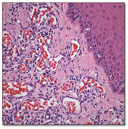
Figure 5. High power (x400) histology shows dilated and congested blood vessels of variable shapes and sizes lined by one layer of flat endothelial cells.
After you have finished reviewing the available diagnostic information