Return to Case of the Month Archives
Relatively Well-Demarcated Radiolucency with Sharply Angulated Tooth Resorption
Dolphine Oda, BDS, MSc
doda@u.washington.edu
Contributed by
Dr. Ashoka Subedar
Bellingham Oral & Maxillofacial Surgery, Bellingham, WA
Case Summary and Diagnostic Information
This is a 42-year-old white male with a one-year history of an aggressive radiolucency of the anterior mandible causing a sharp edge and irregular resorption of teeth #s 23 & 24.
Diagnostic Information Available
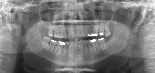
This is a 42-year-old white male with a one-year history of an aggressive radiolucency of the anterior mandible (Figure 1) causing a sharp edge and irregular resorption of teeth #s 23 & 24. The radiolucency was well-demarcated despite the aggressive tooth resorption. The patient was advised to biopsy the area but he did not come until one year later with a larger radiolucency, still well-demarcated but with more significant tooth resorption involving teeth #s 22-25 (Figure 2). The teeth at this point were loose and there was obvious vestibular expansion which was described to be pink/purple in color. The swelling was approximately 2 x 3 cm in size.
Figure 1 This radiograph was taken in 2009 at first clinical presentation. Note the relatively well-demarcated radiolucency anterior mandible but the very sharp resorption of tooth #23 and irregular resorption of tooth #24.
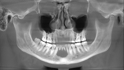
Figure 2 This radiograph was taken in 2010 one year after the first presentation. Note the relatively well-demarcated but larger radiolucency in the anterior mandible with irregular and significant resorption of teeth #s 22-25.
The patient’s past medical history is negative for any significant disease or risk factors.
The patient reported progressive swelling of the anterior mandible over one-year period. The anterior teeth became loose over a one-year period and the swelling increased progressively. The swelling was more in the labial vestibule than the lingual and the tooth resorption was aggressive and involved multiple teeth. All the teeth were vital.
Under local anesthesia, an incisional biopsy was performed and the specimen was submitted for microscopic examination.
Histologic examination reveals multiple pieces of soft tissue composed of a mass of granulation tissue with many giant cells which are haphazardly arranged (Figures 3 & 4). The granulation tissue is loose, cellular and has many small blood vessels. It contains clusters of hemosiderin pigment. The granulation tissue also contains many giant cells of variable shapes and sizes haphazardly arranged some closely associated with small blood vessels.
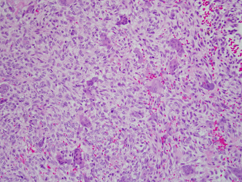
Figure 3 Low power (x100) H & E histology shows a mass of granulation tissue with many giant cells which are haphazardly arranged. The granulation tissue is loose and cellular, with many small blood vessels.
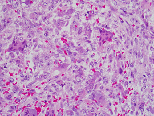
Figure 4 Higher power (x200) H & E histology shows many giant cells suspended on vascular granulation tissue.
After you have finished reviewing the available diagnostic information