Return to Case of the Month Archives
December 2005: Unilocular radiolucency right maxillary sinus
Dolphine Oda, BDS, MSc
doda@u.washington.edu
Contributed by
Dr. Franco Audia, Bellevue-Redmond OMS
Case Summary and Diagnostic Information
This is a 39-year-old white male who was referred by his ENT physician to Dr. Audia after refractory treatment for chronic sinusitis.
Diagnostic Information Available
This is a 39-year-old white male who was referred by his ENT physician to Dr. Audia after refractory treatment for chronic sinusitis with numerous antimicrobial agents including Levaquin and Nasonex. A Computed Tomography (CT) scan (Fig 1) of the right maxillary sinus was performed, revealing a mass in the right maxillary sinus associated with the apices of tooth #2.
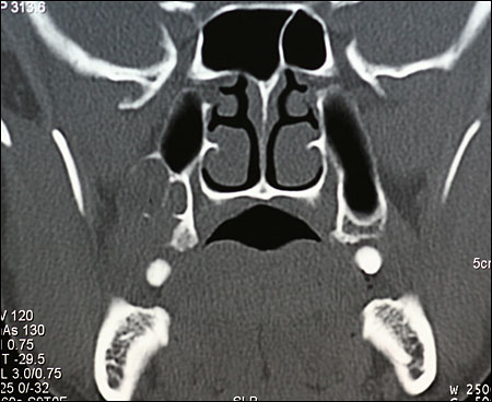
Figure 1.
His medical and family history is non-contributory.
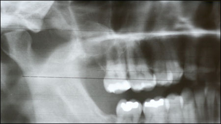
The CT scan (Fig 1) and the Panoramic (Fig 2) images revealed a well-demarcated and corticated unilocular radiolucent lesion in the right posterior maxilla and right maxillary sinus. Clinically, there was evidence of a fistula behind tooth #2. Teeth in the upper right maxilla tested vital and were asymptomatic.
Figure 1. This a coronal computed tomography (CT) scan view at presentation demonstrating a radiolucent area extending to the right tuberosity area and filling half of the right maxillary sinus.
Figure 2. This is a panoramic radiograph taken at the primary presentation demonstrating a well-demarcated and unilocular radiolucency in the posterior maxilla and right maxillary sinus.
An incisional biopsy was performed in September 2005; purulent exudate was noted at the time of the biopsy. The histologic examination of the incisional biopsy revealed a cystic structure lined by thin and corrugated epithelium (Fig 3) and supported by a thin fibrous connective tissue wall. The latter was focally inflamed. The epithelium was keratinized and had a palisaded basal cell layer (Fig 4).
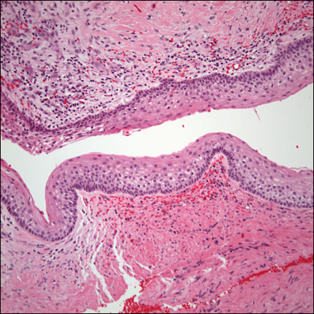
Figure 3. Low power (x100) histology shows a cystic structure lined by a uniformly thin lining epithelium which is supported by a fibrous connective tissue wall. The lining epithelium is corrugated and keratinized.
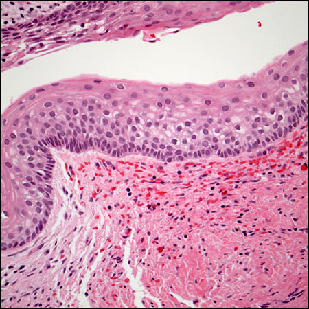
Figure 4. Higher power (x200) histology shows a cystic structure with palisaded basal cell layer.
After you have finished reviewing the available diagnostic information