Return to Case of the Month Archives
July 2010: Blue Hard Palate
Dolphine Oda, BDS, MSc
doda@u.washington.edu
Contributed by
Dr. Todd Carter
Maui Oral & Maxillofacial Surgery, Maui, Hawaii
Case Summary and Diagnostic Information
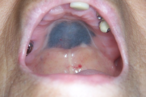
This is a 78-year-old white female who has had a uniformly blue lesion in her hard palate for over two years. It is otherwise asymptomatic and is not present anywhere else.
Diagnostic Information Available
This is a 78-year-old white female who has had a uniformly blue lesion in her hard palate for over two years. It is otherwise asymptomatic and is not present anywhere else. The patient reports long-term use of the following drugs: Plaquenil (20mg) 1-2x/day; Simvastatin (5mg) 1-3x/day; Prednisone (1.4mg) as needed; Plavix (75mg) 1x/day; Protonix (40mg) as needed; Zyrtec (10mg) as needed; Tylenol (regular strength) as needed and Ferrous Sulfate (125mg) 1x/day.
Figure 1 Clinical photograph of the lesion taken at the first visit. Note the flat, uniform color, diffuse blue color involving almost all the hard palate.
This patient’s past medical history is significant for coronary artery disease, hypertension, rheumatoid arthritis, asthma, peptic ulcer and osteoporosis.
This flat, generalized, uniformly blue pigmented lesion affected most of the hard palate but was otherwise asymptomatic. There is no other discoloration anywhere else in her mouth or skin. The lesion was of more than two years’ duration and was not painful, and there was no evidence of a swelling or infection.
Treatment
Under local anesthesia, a small biopsy was taken. The results of the incisional biopsy were such that further treatment was deemed unnecessary.
Incisional Biopsy
Histologic examination of the H & E section revealed a trisected small piece of mucosa covered by epithelium and supported by dense fibrous connective tissue (Figure 2). The connective tissue contained small clusters of a dark-brown granular material, some of which was golden brown and finely granular which stained positively for melanin stain Masson Fontana (Figure 3). There were other small clusters of brown pigment that was larger and more variable in size dark brown granules that stained blue with iron stain (Figure 4). Figure 4 shows a combination of dark blue pigment consistent with hemosiderin and small granular golden-brown pigment consistent with melanin. Immunohistochemistry stain was negative with S-100 antibody.

Figure 2 Low power (x100) histology shows H & E stained section with surface epithelium and underlying dense fibrous connective tissue containing small clusters of brownish granular material.

Figure 3 Higher power (x200) histology shows Masson-Fontana, special stain for melanin demonstrating small clusters of brown pigment staining positively with Masson Fontana. The pigment is golden brown and finely granular.

Figure 4 Higher power (x200) histology shows iron stain for hemosiderin pigment. Note the clusters of blue pigment staining positively with iron stain confirming that some of the brown pigment was hemosiderin.
After you have finished reviewing the available diagnostic information
