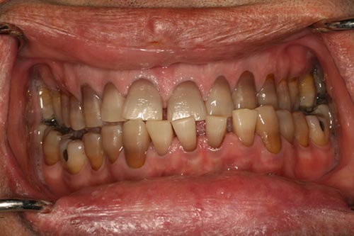
Enlarging mandible, spaced teeth, large tongue and coarse facial appearance
Can you make the correct diagnosis?
This is a 44-year-old white male who presented to Dr. Snyder as a new patient. His main complaint was that he had an under bite and that his “lower front teeth seem to be drifting and moving anteriorly” (Figure 1). He had a mountain bike accident the year before. His brother is a physician who lives in a different city had recently commented to his brother that his face “seems different.” On examination, he had a bilateral class III bite with cross bite on the left side and edge to edge on the right side. His tongue was very large and his lips (especially the lower lip) were full and had frontal bossing (Figure 2). The panoramic radiograph was not large enough to show his entire mandible A-P and left to right. His lower teeth were spaced and all of his teeth were internally dark purple in color. On further questioning, the patient also stated that his hands and feet were growing and that his gloves “keep getting too small.”
Congratulations! You are correct
Acromegaly, a rare disease, is slow and progressive and is often not recognized for years (1-4). There is an average of 7-10 years’ delay in diagnosis. Given the tendency of this disease to affect small and facial bones, especially the mandible, it would be reasonable to assume that a primary dental practitioner would be the first to recognize the disease in many cases.
Acromegaly is caused by an overproduction of growth hormone (GH) by the pituitary gland due to either glandular hyperplasia, benign adenoma or a malignant neoplasm (2-4). Pituitary functional adenomas are usually the most commonly associated with this disease. Pituitary adenomas are reported in about 10% of the normal population and are classified into those that produce excessive hormones (i.e. prolactin, GH, corticotrophin) or those that are non-hormone producing. Approximately 20% of the functional pituitary adenomas are GH producing (1-2). Excessive GH production causes acromegaly in adults and gigantism in children. It affects males and females equally, usually around the age of 40-45 (3-4).
The symptoms associated with acromegaly are the result of either overproduction by the pituitary adenoma or underproduction of the pituitary gland due to the adenoma replacing the normal pituitary gland by tumor mass (1-3). The symptoms associated with GH overproduction are numerous and range from cardiovascular diseases to bone overgrowth, usually of facial bones and the small bones of the hands and feet. Facial bone changes include frontal bossing, enlarging mandible leading to prognathism, tooth spacing, apical root resorption (usually mandibular molars and maxillary central incisors) and an enlarging nose, leading to a coarse facial appearance (1-4). Soft tissue facial changes include a full lower lip, macroglossia and swelling of the soft palate leading to sleep apnea. Acromegaly almost always affects the small bones of the hands and feet, leading to large and square hands. Other symptoms include cardiovascular diseases such as hypertension, congestive heart failure, diabetes mellitus, fatigue and osteoarthritis (2-3).
Patients may seek dental help because of the enlarging mandible/prognathism and, if edentulous, they may complain of ill-fitting dentures. The patients may also complain of ill-fitting shoes and hats, headaches, and/or visual defects due to the tumor expansion (1-3).
Laboratory tests include measurement of serum growth hormone in response to glucose load and pituitary gland imaging (such as MRI) (1-4). Treatment, ranging from surgery to radiation therapy, depends on the size of the neoplasm. Treatment should also include controlling the serum GH level by one of the somatostatins or via the GH receptor-blocking agents (2-3). The prognosis depends on when the disease is discovered and the number of systems involved. Corrective jaw surgery can be helpful in resuming normal occlusal alignment and function.
References
- Hurley DM, Ho KKY, Pituitary disease in adults, Med J Aust 180 (2004), pp. 419–425.
- Gsponer J, DeTribolet N, Deruaz JP, Janzer R, Uske A, Mirimanoff ROet al., Diagnosis, treatment, and outcome of pituitary tumors and other abnormal intrasellar masses Retrospective analysis of 353 patients, Medicine 78 (1999), pp. 236–269.
- Melmed S. Acromegaly, N Engl J Med 322 (1990), pp. 966–977.
- Laws, ER, Vance ML and Thapar K, Pituitary surgery for the management of acromegaly, Horm Res 53 (2000), pp. 71–75.
- Parekh SG, Donthineni-Rao R et al. Fibrous Dysplasia. J Am Acad Orthop Surg. 2004;12:305-313.
- Tsai EC, Santoreneos S et al. Tumors of the skull base in children: review of tumor types and management strategies. Neurosurg Focus. 2002: 15;12(5).
- Zacharin M. Paediatric management of endocrine complications in McCune-Albright syndrome. J Pediatr Endocrinol Metab. 2005;18:33-41.
- Martinez-Lage JL, Almeida F, Picón M, Lorenzo F, Carrillo R. Maxillomalar monoblock removal, reshaping, and reinsertion in Paget’s disease: 15-year follow-up. J Oral Maxillofac Surg. 2005 Nov;63(11):1680-5.
- Rasmussen JM, Hopfensperger ML. Placement and restoration of dental implants in a patient with Paget’s disease in remission: literature review and clinical report. J Prosthodont. 2008 Jan;17(1):35-40. Epub 2007 Oct 8
Sorry! you are incorrect
With the exception of the enlarging mandible, the other clinical presentations, including the age of this patient (44), are not supportive of fibrous dysplasia. Craniofacial fibrous dysplasia can include generalized swelling, but that is exceptionally rare at this patient’s age.
The etiology of fibrous dysplasia is unknown. Monostotic (affecting one bone or one area) FD constitutes approximately 80% of all fibrous dysplasia cases while the polyostotic (affecting multiple bones or multiple areas) constitutes the other 20% (5-7). All fibrous dysplasias tend to be expansile and disfiguring lesions, whether single or multiple. Monostotic FD, which involves the jaws, affects males and females equally (5). It occurs in childhood and at puberty and usually stops growing at age 30. It appears as an asymptomatic swelling of the maxilla or mandible; the maxillary lesion is the most common. It may involve bones other than the maxilla, including the zygoma, sphenoid and others (7). It is usually unilateral and is known to displace teeth, but otherwise is firmly seated. FD is a slow-growing lesion, but rapid growth has been described, especially during puberty (6-7). The radiographic appearance, especially of the maxilla, is classically described as a ground glass appearance where fine radiopacity is noted. FDs of the mandible lesions are much more deceptive because they tend to vary more, thus making it difficult to diagnose based on the radiograph alone. Mandibular FDs range in radiographic presentation from cystic unilocular radiolucency to multilocular radiolucency to the classical ground glass radio-opacity (5-7).
Polyostotic fibrous dysplasia constitutes approximately 20% of FD cases. Three sub-types are described: craniofacial, Jaffe’s syndrome and Albright’s syndrome. The latter represents the more severe form with endocrine disturbances (precocious puberty in females) (5-7). The Jaffe’s and Albright’s forms of polyostotic FD are more common in females than males. Craniofacial FD constitutes a significant number of polyostotic FDs; these can involve the jaws, zygoma, orbital bone, sphenoids, and frontal bone, among other bones. They are expansile and disfiguring (6). In Jaffe’s and Albright’s cases, multiple bones are affected, including long bones in addition to the jawbones. The clinical presentation depends on the type. Albright’s is associated with hormonal changes, including precocious puberty and cafe au lait spots. Jaffe’s is associated multiple bones with FD and cafe au lait spots but no hormonal changes (5-7). Treatment may be necessary and is preferably performed after cessation of growth due to the high incidence of re-growth and requirement of secondary procedures. Radiation therapy is contraindicated since significant incidence of development of osteosarcoma in the irradiated bone has been documented. Malignancies such as osteosarcoma have been known to arise in areas of FD that have not been irradiated, but rarely (5-7).
Sorry! you are incorrect
The enlarging mandible and tooth spacing are consistent with Paget’s disease. Paget’s disease is more common in the middle face, including maxilla, than the mandible. In addition, the other changes—including full lips, large tongue and hands—are not supportive of this disease.
Paget’s is a sclerosing bone disease of unknown etiology. It can be inflammatory, vascular, or viral, with some work implicating EBV; it may be a prion disease. It consists of three phases: bone resorption, vascular and sclerosing. It typically occurs after the age of 40 (8-9). It is more common in men and more common in the U.K. than in the U.S.A. It is often low-grade and discovered in apparently normal elderly people during autopsy. In the head and neck, the cranium and maxilla are affected more frequently than the mandible (8-9). Paget’s is a chronic, slowly developing disease causing pain, headache, deafness, blindness, facial paralysis and other neural symptoms. The enlarging bone impinges on peripheral nerves. The enlarging maxilla or mandible may result in dentures no longer fitting or the development of spacing in the dental arch. Separation of teeth and hypercementosis are common. Radiographically, it initially appears as an osteolytic lesion (‘ground glass’ appearance) progressing to irregular sclerotic masses with a ‘cotton-wool’ appearance. Softening of bones leads to bowing of the legs and development of a ‘simian gait.’ Laboratory findings: These include a significant increase in serum alkaline phosphatase. Paget’s patients also have an increased incidence of developing osteosarcoma. Therapy is directed toward reducing bone turnover (8-9). Bisphosphonates such as etidronate have been used with success. Other agents sometimes useful include calcitonin and cytotoxic drugs. Surgical recontouring is not indicated due to the increased incidence of postoperative osteomyelitis in the dense, poorly vascular bone. Prognosis depends on the extent of the disease.
