Ill-defined radiolucency, anterior mandible
Can you make the correct diagnosis?
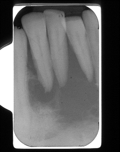
This is a 58-year-old male who was referred by his dental hygienist for evaluation of an anterior mandible midline bony lesion that had been present for several months.
Sorry! you are incorrect
The location of anterior mandible crossing the midline and the radiographic presentation of radiolucency are compatible with some aspects of CGCG. However, the age of the patient is not and the ill-defined radiolucency is not typical of CGCG. The histology, however, is not supportive of CGCG.
Jaffe first coined the term “reparative” for central giant cell granuloma. Most pathologists have since dropped the term “reparative” for lack of evidence that the pathogenesis is a reparative process. CGCG is described as a non-neoplastic process and yet can behave in a very aggressive and expansile manner, destroying bone and displacing teeth. Over 60% of CGCG cases occur in patients younger than 30 years of age, with twice as many occurrences in females as in males. CGCG is classified into aggressive and non-aggressive types; the aggressive type tends to occur in younger patients and causes disfiguration, especially after surgery. Over 70% of cases occur in the mandible anterior to the first molar tooth. This lesion has also been described in other cranio-facial and small long bones such as those of the hands and feet.
The usual treatment for CGCG is surgery, ranging from curettage and en bloc to resection. The latter is used in aggressive and recurring cases. In the past ten years or so, alternatives to surgery have emerged with successful results, saving some patients from facial disfigurement. Steroid injections are the most successful alternative treatment thus far; they require injections weekly or every 2-3 weeks, have no known side effects (even in children), and are the least expensive alternative treatment. Other treatments include: calcitonin injections or nasal spray, which require daily injections or a nasal spray of salmon calcitonin for about a year and are safe for pregnant females; and interferon alfa-2a injections, which are administered 2-3 times per week for several months and are the most expensive alternative treatment.
Sorry! you are incorrect
Again, the ill-defined radiolucency should make one think aggressive behavior and may be even malignant behavior such as primary malignancy or metastatic disease. Anterior mandible is not a common location for metastatic disease and for that matter is not a common location for primary malignant neoplasms with the exception of chondrosarcoma. The latter would have been considered on the differential diagnosis if the lesion was mixed radiolucent/radiopaque or predominantly radiopaque. It is highly unusual for chondrosarcoma to be all radiolucent. Peripheral blood malignant neoplasms are usually completely radiolucent and can occur in the jaw bones, especially in the mandible but more in the posterior mandible. One example or peripheral blood malignancy to consider would be plasmacytoma, a malignant neoplasm of plasma cell origin. The age and gender of this patient is good for plasmacytoma, so are the radiographic changes and the 3+ mobility of the involved teeth. The histology however is not supportive of this disease.
Plasmacytoma is an isolated manifestation of multiple myeloma. Plamacytoma or multiple myeloma is a clonal neoplastic proliferation of plasma cells (myeloma cells) which are terminally differentiated B-lymphocytes. It is most common in individuals 60-75 years of age, occurs more in males, and is rare in persons under 40 years of age. It is twice as common in blacks as it is in whites In the US. It occurs most commonly in bone and in a multicentric manner (multiple locations at the same time), hence the name multiple myeloma. This malignant neoplasm presents more commonly in bone but is also described in soft tissue without bony involvement (extramedullary). About 40-50% of bone-isolated plasmacytomas will progress to multiple myeloma while only 10-12% of soft tissue plasmacytomas will progress to multiple myeloma. Normal plasma cells are immunoglobulin producing cells including IgA, IgD, IgG, IgM, and IgE. Neoplastic plasma cells tend to produce any of these antibodies, but predominantly IgG and IgA. The most common clinical presentation is severe bone pain, especially that of the lumbar spine which is usually aggravated with movement. Bone lesions occur in 70% of patients and usually lead to hypercalcemia, bone fracture and vertebral collapse. Hypercalcemia may lead to soft tissue calcifications. The most common bones affected by multiple myeloma are the vertebrae, skull, mandible, ribs, pelvis, clavicles and scapula. Jaws are affected in about 30% of cases. Extramedullary tissue such as the liver, spleen and lymph nodes can also be affected. Patients with multiple myeloma are susceptible to anemia and therefore often experience fatigue and weakness. They can also develop renal failure due to monoclonal light chain (Bence Jones protein). About 15% of patients develop amyloid deposition in a variety of soft tissues, including the G-I tract and the oral cavity, specifically the tongue. Amyloid in the tongue can present as small yellow nodules or plaques; they can be deep seated within the tongue, causing slurring. They can also occur around the eyes, again as small yellow plaques or nodules. Radiographically, multiple myeloma has a distinct presentation of “punched out” radiolucencies but the radiolucency can also be ill defined. This is particularly true of the skull lesion. The mandible and maxilla can be affected, but the radiographic presentation is not as distinct as in the case of skull lesions. It is important to note that histology and immunohistochemistry alone cannot differentiate between an isolated case of plasmacytoma and the full disease of multiple myeloma. Pathologists usually render the histologic diagnosis of plasmacytoma while oncologists render the clinical diagnosis of multiple myeloma.
Histologically, the biopsy of a patient with isolated plasmacytoma or a patient with the full disease of multiple myeloma demonstrates sheets of monotonous mononuclear cells with plasmacytic differentiation. The atypical plasma cells usually show nuclear and cellular atypia. Mitosis is usually present. The neoplastic cells are monoclonal; this can be supported by an immunohistochemistry stain with kappa or lambda light chains. They are also specific with CD138, a marker for plasmacytoid cell. The final diagnosis of multiple myeloma is determined through a combination of histology, serum and urine protein electrophoresis, blood test for myeloma protein (monoclonal gammopathy), and radiographic changes. Treatment includes chemotherapy, bisphosphonates to control hypercalcemia, bone marrow transplant and radiation therapy. The overall prognosis depends on age; younger patients have a better prognosis than older patients.
Sorry! you are incorrect
The ill-defined radiolucency and destructive nature of the disease especially, the 3+ mobility of the anterior teeth makes LCH a valid condition to be included on the differential diagnosis. The age of the patient however is not supportive of this condition since LCH tends to occur in children and young adults. The location of anterior mandible is not typical of LCH. The histology is also not supportive of LCH.
Langerhans cells are dendritic cells of bone-marrow origin. Langerhans cell histiocytosis is generally a disease of children, only rarely affecting adults. The old name for this condition was histiocytosis X, which was classified into eosinophilic granuloma (monostotic and polyostotic), Hand-Schuller-Christian disease, and Letterer-Siwe disease. The first two types (EG and HSC) are less aggressive and are chronic in clinical behavior while the last (LS) is more aggressive and acute in behavior. The currently used classification divides cases into the categories of unifocal, multifocal/unisystem and multifocal/multisystem. This classification also divides patients into low- and high-risk categories: patients with the unifocal and multifocal/unisystem types are considered low risk, while those with the multifocal/multisystem type are high risk. In other words, if the disease affects one organ, whether in one or more sites (i.e. bone, lymph nodes, skin or pituitary gland), it is considered to be a low-risk type; if the disease involves multiple organs such as the lungs, liver, spleen and bone marrow (multifocal and multisystem), it is considered to be a high-risk type. LCH usually presents with pain and swelling but can also be asymptomatic. The bony lesions are usually aggressively lytic, especially those of the mandible. More than 50% of cases affect children under 10 years of age, with a male predilection. Adult LCH most commonly affects the bones, especially the jaws and skull bones in general. LCH of the jaw bone usually presents as either localized severe periodontitis or generalized severe periodontitis, and rarely as localized, well-demarcated radiolucency. Radiographically, the bony lesions are sharply radiolucent and the jaw lesions give the impression of teeth “floating” in space due to the significant bone destruction. LCH can also affect the skin, the lymph nodes, and less commonly the pituitary and lungs.
Langerhans cells are CD1a positive and for that reason, immunohistochemistry staining is very helpful. They are also S-100 and CD45 positive. Langerhans cells can also be comfortably identified by the H & E stain. Treatment includes curettage especially of the jaw lesions. Chemotherapy and low-level radiotherapy have also been effective. The unifocal disease has an overall 95% survival rate, types with two-organ involvement have an overall survival rate of 75%, and the more organs are involved, the less favorable the prognosis.
Congratulations! You are correct
Around 60,000 cases of non-Hodgkin’s lymphomas are reported annually in the U.S.A. and about one third die of the disease. Diffuse large B cell lymphoma (DLBCL) and follicular lymphoma (FL) are the most common types with DLBCL being more common and more aggressive in behavior. Non-Hodgkin’s lymphoma is a malignant neoplasm of lymphocyte origin, mostly of B-lymphocyte but can also be of T-lymphocyte origin. Several of skin lymphomas are of T-lymphocyte origin. The head and neck region is a frequent site for lymphomas, especially the Waldeyer’s ring. However, the oral cavity is not a common site except in certain types such as African type Burkitt’s lymphoma. This malignant lymphoma involved the jaw bone as the primary lesion. Primary non-Hodgkin lymphomas of bone are rare and constitute around 2% of all adult lymphomas and up to 9% of children’s lymphomas. They are slightly more common in males and are more common in individuals over the age of 60. Clinical presentation includes pain, swelling, fracture of the involved bone. Malignant lymphomas of the jaws also present in form of severe periodontitis and bone loss with tooth mobility as is the case in this patient. In some cases, the symptoms may be present for months as was the case in this patient even mimicking infection which is another manifestation of this disease. Primary lymphoma of bone is staged in the same way as any patient with non-Hodgkin lymphoma involving the lymph nodes and other soft tissues. The Ann Arbor staging classification is usually used and a single disease area confined to one bone would be staged low and if involving multiple sites and multiple bones would be staged as IV. Treatment for primary lymphoma of bone is radiation therapy with good results. However, radiation alone has not be successful in terms of recurrence in sites away from the primary lesions and for that reason, chemotherapy is currently used with and without radiation therapy.
References
- Whitaker SB, Vigneswaran N, Budnick SD, Waldron CA. Giant cell lesions of the jaws: evaluation of nucleolar organizer regions of varying behavior. J Oral Pathol Med 1993; 22(9):402-5.
- Tallan EM, Olsen KD, McCaffrey TV, Unni KK, Lund BA. Advanced giant cell granuloma: a twenty-year study. Otolaryngol Head Neck Surg 1994;110:413-8.
- O’Regan EM, Gibb DH, Odell EW. Rapid growth of giant cell granuloma in pregnancy treated with calcitonin. Oral Surg Oral Med Oral Pathol Oral Radiol Endod 200; 92(5):532-8.
- Collins A. Experience with anti-angiogenic therapy of giant cell granuloma of the facial bones. Ann R Australas Coll Dent Surg 2000; 15:170-5.
- Uhler IV, Fahs GR, Dolan LA. Metastasis of cervical carcinoma to the mandible: report of a case. J Am Dent. Assoc. 1972; 85: 363-364.
- Praetorius F, Hjorting-Hansen E, Gorlin RJ, Vickers RA. Calcifying odontogenic cyst: range, variations and neoplastic potential. Acta Odonotol Scand 1981;39:227-240.
- Akhtar K, Laghari NA, Haq AU, Anees M, Rehman SU, Alam MI. Multiple myeloma in younger age.. J Coll Physicians Surg Pak. 2009 Jan;19(1):62-3.
- Poggio CE. Plasmacytoma of the mandible associated with a dental implant failure: a clinical report. Clin Oral Implants Res. 2007 Aug;18(4):540-3. Epub 2007 Apr 30.
- Canger EM, Celenk P, Alkan A, Günhan O. Mandibular involvement of solitary plasmocytoma: a case report. Med Oral Patol Oral Cir Bucal. 2007 Jan 1;12(1):E7-9.
- Bonet J, Manuel MJ et al. Eosinophilic granuloma of the jaws: a report of three cases. Med. Oral. 2001; 6: 218-224.
- Favara BE, Feller AC, Pauli M et al. Contemporary classification of histiocytic disorders. Med Pediatr Oncol 1997; 29: 157–66.
- Baumgartner I, von Hochstetter A, Baumert B et al. Langerhans cell histiocytosis in adults. Med Pediatr Oncol 1997; 28: 9–14.
- Malpas JS, Norton AJ. Langerhans cell histiocytosis in the adult. Med Ped Oncol 1996; 27: 540–6.
- Epstein JB, Epstein JD, Le ND, Gorsky M. Characteristics of oral and paraoral malignant lymphoma: a population-based review of 361 cases. Oral Surg Oral Med Oral Pathol Oral Radiol Endod 2001, 92:519-525.
- Urquhart A, Berg R. Hodgkin’s and non-Hodgkin’s lymphoma of the head and neck. Laryngoscope 2001, 111:1565-1569.