October 2010: Recurring Lobular Exophytic Lesion Right Lateral Tongue
Can you make the correct diagnosis?
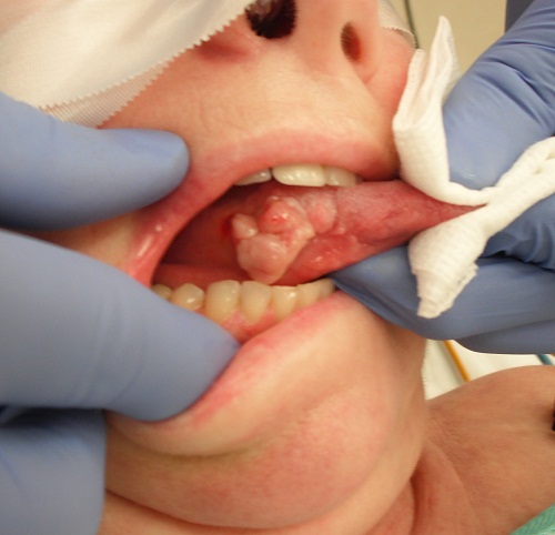
This is a 49-year-old white female who was referred to the University of Washington, Department of Oral Surgery for a fast-growing, recurring, exophytic mass on the right lateral border of tongue present since August 2008.
Sorry! you are incorrect
The lobular exophytic swelling on the lateral/ventral tongue with a history of trauma should make one think of pyogenic granuloma or a reactive fibroblastic-type lesion. The histology in this case, however, is not consistent with PG.
Pyogenic granuloma constitutes 85% of all reactive gingival swellings but can occur in other areas such as the tongue. Pyogenic granuloma is a mass of exuberant vascular granulation tissue (1). It can be induced by local irritants such as a sharp bite, sharp fillings, excessive plaque and dental calculus. Tt sometimes forms in an extraction socket in response to an irritant left in the socket. The gingiva are the most common location, but it can occur anywhere in the oral cavity and skin, especially the tongue, lips, fingers and nail beds (1). In the mouth, it occurs most commonly in the gingiva, especially the maxillary buccal and interproximal gingiva (1-2). Occasionally, it may surround a tooth. It is usually highly vascular, fast-growing, exophytic, lobular, sessile, and ulcerated or covered by pseudomembrane. The color changes from red to pink when it starts to heal. It occurs at any age and sex with a slight predilection for young females; it affects 1% of pregnant females. Pyogenic granuloma is usually painless except during eating, when bleeding and pain are described (1). Histologically, it presents as a mass of loose and vascular granulation tissue, usually with ulcerated or eroded surface epithelium and many inflammatory cells. A range of treatment modalities are available, including excision with removal of the local irritant, laser surgery, or intralesional injection with absolute alcohol, steroids or botulinum toxin (2-3). Elimination of the causative local stimulus prior to surgical removal helps shrink the lesion. The prognosis is good, although recurrence is possible, especially during pregnancy.
Sorry! you are incorrect
Due to the location of ventral/lateral tongue in this case, there is a high-risk possibility of oral squamous cell carcinoma. The location, when combined with history of recurrence, should make the clinician think of SCC and request an incisional biopsy to rule it out. The histology in this case is not supportive of oral SCC.
Squamous cell carcinoma of the mouth is a highly aggressive neoplasm that currently ranks as the fifth most common malignant neoplasm worldwide and accounts for an estimated 90% of oral malignancies (4). Oral SCC occurs predominantly in males over the age of 40 years, with an observed male-to-female ratio of 2:1 generally and 1.4:1 in the USA (4-6). Excluding the outer lip, the most common sites (in decreasing order) are the ventral and lateral surfaces of the tongue (25-50%), floor of mouth (15%), gingiva (12%) and palate (9%). The buccal mucosa and retromolar pad areas (3%) have a relatively low incidence of occurrence (4-5) unless the patient is a chronic smokeless tobacco user. Oral SCC varies in presentation from deceptively innocent-looking to obviously malignant. It may present as a non-healing ulcer, or as red, white or mixed red-and-white lesions. Characteristic signs of oral SCC are non-healing ulcer, ulcer with rolled borders, fungation, fixation and induration. Rarely, oral SCC may present as an unexplained asymptomatic lateral neck lymphadenopathy (4-6). Oral SCC is most commonly associated with chemically induced mutagenesis, specifically tobacco and alcohol use (4-5).
Tobacco use is described in over 75% of oral SCC patients (4-5).Tobacco and alcohol have been shown to act synergistically in the development of oral SCC. Human papilloma virus (HPV) has also been found to have a high prevalence in oral cancer, especially in younger patients with no history of tobacco use (6). Other factors include poor oral hygiene, syphilis, chronic candidiasis, iron and dietary deficiencies, herpes simplex and various other immunologic factors, and lichen planus—especially persistent erosive lichen planus.
Determination of the prognosis of oral SCC is based on its clinical stage and histological classification (4-6). Although oral SCC is a diagnosis made by histology, surgeons tend to depend exclusively on the TNM classification system for clinical staging and treatment decisions. Prognosis is dependent on the TNM staging system; the most important prognostic sign is the presence or absence of metastases at the time of diagnosis. The prognosis thus improves when the lesion is detected early. Oral SCC patients die mainly of infection due to lowered resistance or of hemorrhage if the tumor erodes through one of the main blood vessels.
Congratulations! You are correct
Traumatic ulcerative granuloma with stromal eosinophilia is known by a number of other names, including traumatic granuloma, eosinophilic ulcer and eosinophilic granuloma of the tongue (7-9). (This condition is not related to eosinophilic granuloma of the bone as in Langerhan cell histiocytosis.) This is a reactive, self limiting condition of the oral cavity. It occurs most commonly by far on the dorsal and lateral tongue, followed by the lips and buccal mucosa (7-9). Acute trauma in the form of a sharp puncture to the muscle is the main cause, but occasionally a history of trauma is absent and the etiology is unknown (7-9). The source of trauma can be a sharp tooth, a sharp filling, an ill-fitting partial denture, or a physical sharp bite (which may or not be due to a neurological disorder). Dorsal tongue TUGSE is usually due to trauma from the maxillary incisors. TUGSE may affect patients of all ages, including infants (8). In infants, it is known as Riga-Fede disease, which occurs during the first week to first year of life (8). Rege-Fede is histologically similar to TUGSE and is the result of trauma, usually to the anterior ventral surface of tongue, followed in frequency by the dorsal tongue surface. It is usually induced by the baby’s mandibular or maxillary natal or prenatal sharp-edged incisors during breastfeeding (7-8). However, TUGSE is more common in adults over the age of forty and more common in males (7) with a male-to-female ratio of 1.6:1. The clinical presentation varies from deep, rolled bordered and indurated to exophytic and lobular looking like a pyogenic granuloma (7-9). The deep and indurated morphology is frequently mistaken for invasive squamous cell carcinoma (SCC). Another aspect of the clinical behavior that may simulate oral SCC is the tendency of TUGSE to recur and to heal slowly; both are reasons to suspect SCC. Traumatic ulcers of the mouth are common and tend to heal within ten days to two weeks while TUGSE may last for many weeks. It is important to know that single deep ulcers of the mouth should be biopsied with two to three weeks of occurrence. Sometimes, an incisional biopsy helps the area to granulate and heal. Histologically, these ulcers are deep lesions involving the underlying muscle, which may explain the process of slow healing and the tissue eosinophilia. Healing may take up to eight weeks. Eosinophils are found in areas of muscle damage. The treatment of choice is surgery but elimination of the causative factor should come first, such as the filing of sharp teeth, the replacement of broken fillings, the use of a thin mouthguard, or whatever else is necessary. Conservative surgical removal with clean margins is the treatment of choice. Sometimes, an incisional biopsy may lead to complete recovery, other times; it may recur and require further surgery with clean margins (7-9). Intra-lesional corticosteroid injections have been recommended for recurring ulcers (7) but with a mixed outcome. The overall prognosis is good.
References
- Fantasia JE, Damm DD. Red nodular lesion of tongue. Pyogenic granuloma.
Gen Dent. 2003 Mar-Apr;51(2):190-194. - Ichimiya M, Yoshikawa Y, Hamamoto Y, Muto M. Successful treatment of pyogenic granuloma with injection of absolute ethanol. J Dermatol. 2004 Apr;31(4):342-4.
- Pham J, Yin S, Morgan M, Stucker F, Nathan CA. Botulinum toxin: helpful adjunct to early resolution of laryngeal granulomas. J Laryngol Otol. 2004 Oct;118(10):781-5.
- Bundgaard T, S Bentzen, et al. Histopathologic, stereologic, Epidemiologic, and clinical parameters in the prognostic evaluation of squamous cell carcinoma of the oral cavity. Head & Neck. 18:142-152 (1996).
- Barasch A, DE Morse, et al. Smoking, gender, and age as risk factors for site-specific intraoral squamous cell carcinoma. Cancer 73:509-513 (1994).
- Syrjanen SM, KJ Syrjanen et al. Human papillomavirus (HPV) DNA sequences in oral precancerous lesions and squamous cell carcinoma demonstrated by in situ hybridization. J Oral Pathol. 17:273 (1988).
- Neville BW, Damm DD, Allen CM, Bouquot JE. Traumatic ulcerations. In: Oral and Maxillofacial Pathology, 3rd edition. Philadelphia: W.B. Saunders, 2009. p. 287-289.
- Richard PE. Richmond VA: Traumatic ulcerative granuloma with stromal eosinophilia (Riga-Fede’s disease and traumatic eosinophilic granuloma), Oral Surg, Oral Med. Oral Pathol, Endod, 1983, vol 55: 497-506.
- Sklavounou A, Laskaris G: Eosinophilic ulcer of oral mucosa, J Oral Surg, 1998, vol 58: 431 – 436.