November 2009: Unilocular radiolucency around impacted tooth #22
Can you make the correct diagnosis?
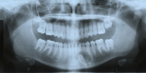
This is an 11-year-old white female who was referred by her orthodontist for evaluation of a “cyst” associated with impacted tooth #22 (Figure 1).
Sorry! you are incorrect
When examining a unilocular radiolucency around the crown of an impacted tooth, the clinician should first consider the possibility that it is a dentigerous cyst, followed by other possibilities in a differential diagnosis. Accordingly, the radiographic presentation of this case is typical of a dentigerous cyst. However, given the location of this lesion (anterior mandible), as well as the age and gender of this patient (a young female), other lesions should be seriously considered. The histology in this case is not supportive of a dentigerous cyst.
Dentigerous cyst is the most common developmental cyst in the oral cavity, accounting for 20% of the developmental cysts of the jaws, and is almost always associated with the crown of a tooth attached to the cemento-enamel junction as is the case in this patient. It is believed to originate from the accumulation of fluid between the reduced enamel epithelium and the tooth crown, thus expanding the follicle beyond the 3 mm normal diameter (1-3). It is usually associated with impacted or un-erupted teeth. Clinically, it can be an asymptomatic radiolucency discovered during a routine dental examination or can act aggressively by expanding the jaws, leading to facial asymmetry with extreme displacement of adjacent teeth as well as the involved tooth (1-2). It can be painful and cause root resorption, especially if it is infected. It is more common in the third molars and upper canines and can also involve supernumerary teeth and odontomas. Radiographically, it presents as a smooth border, usually of unilocular or sometimes multilocular radiolucency, located around the tooth circumferentially, laterally or in the form of a “doughnut” ring. The size varies from small to extremely large, extending into the ramus of the mandible. The borders are usually corticated and smooth unless it is inflamed when loss or cortication (usually partial) is present (1). Histologically, the cyst has non-specific features lined by variable thickness of stratified squamous epithelium and supported by connective tissue. Clusters of mucous cells are frequently present, especially in the mandibular third molar areas. Treatment depends on its size, and ranges from thorough curettage to marsupialization. It usually has a good prognosis. Recurrence is uncommon if properly removed. Occasionally this cyst is associated with ameloblastoma, epithelial dysplasia and/or squamous cell carcinoma or mucoepidermoid carcinoma (1-3).
Tobacco use is described in over 75% of oral SCC patients (7-9). Tobacco and alcohol have been shown to act synergistically in the development of oral SCC. Human papilloma virus (HPV) has also been found to have a high prevalence in oral cancer, especially in younger patients with no history of tobacco use. Other factors include poor oral hygiene, syphilis, chronic candidiasis, iron and dietary deficiencies, herpes simplex and various other immunologic factors, and lichen planus—especially persistent erosive lichen planus.
Determination of the prognosis of oral SCC is based on its clinical stage and histological classification (7-9). Although oral SCC is a diagnosis made by histology, surgeons tend to depend exclusively on the TNM classification system for clinical staging and treatment decisions. Prognosis is dependent on the TNM staging system; the most important prognostic sign is the presence or absence of metastases at the time of diagnosis. The prognosis thus improves when the lesion is detected early. Oral SCC patients die mainly of infection due to lowered resistance or of hemorrhage if the tumor erodes through one of the main blood vessels.
Sorry! you are incorrect
A unilocular radiolucency around an impacted tooth should make the clinician consider the possibility that it is an odontogenic keratocyst; this possibility is second to that of it being a dentigerous cyst. Both the clinical and the radiographic findings of this case are supportive of an OKC but the histology is not.
Odontogenic keratocyst (OKC) is an aggressive odontogenic cyst and is known for its rapid growth and its tendency to invade the adjacent tissues, including bone. It has a high recurrence rate and is associated with basal cell nevus syndrome (4-5). The majority of patients are in the age ranges of 20-29 and 40-59, but cases in patients ranging in age from 5 to 80 years have been reported (4). The distribution between sexes varies from equal distribution to a male-to-female ratio of 1.6:1. OKC predominantly affects Caucasian populations and, if one may judge from the limited evidence provided by the literature, is chiefly of Northern European descent (4). Odontogenic keratocysts may occur in any part of the upper and lower jaw, with the majority (almost 70%) occurring in the mandible. They occur most commonly in the angle of the mandible extending superiorly into the ramus (4-5). Radiographically, OKCs present predominantly as unilocular radiolucencies with well-defined or sclerotic borders; they may also present as multilocular radiolucencies or, more commonly, unilocular with scalloped borders (4-5). They usually penetrate the bone rather than expand; they grow in an anterior and posterior manner with little to no expansion. The larger OKCs, however, tend to expand bone, but mildly. Obvious clinical expansion should be viewed with suspicion for a neoplasm. OKCs can also present as small and oval radiolucencies between teeth simulating a lateral periodontal cyst, in an area of an extracted tooth simulating a residual cyst, at the apex of a vital tooth mistaken for a periapical cyst, or in the anterior maxilla between the central incisors simulating an incisive canal cyst (4-5). OKCs grow to sizes larger than any other odontogenic cysts. Despite this aggressive growth, they often remain asymptomatic, thus growing to large sizes and hollowing the bone (4-5). Like dentigerous cysts, if infected, they can be painful, thus symptomatic. Multiple OKCs are frequently associated with bifid-rib basal cell nevus syndrome (Gorlin syndrome). Odontogenic keratocysts are significant clinical entities due to their tendency for recurrence and destructive behavior. They are known to have a high recurrence rate, ranging from 13% to 60% (4). Complete surgical removal is the treatment of choice. Enucleation combined with Carnoy’s solution or liquid nitrogen treatment has been effective in reducing recurrence rate (5).
Congratulations! You are correct
Adenomatoid odontogenic tumor (AOT) is a rare neoplasm (hamartoma) accounting for less than 3% of all odontogenic tumors. It is benign and slow-growing and is believed to originate from the remnants of the dental lamina or enamel organ. It was first described by Dreibladt, in 1907, as a pseudoadenoameloblastoma (9). In 1969, Philipsen et al proposed the name adenomatoid odontogenic tumor, which was later adopted by the World Health Organization with the understanding that this lesion has a benign and non-aggressive behavior (10). It presents within bone (intra-osseous) in 97.2% of cases and on the gingiva (extra-osseous, also known as peripheral) in 2.8% of cases (11). The intra-osseous type is further divided into follicular (most commonly) and extra-follicular types (11). The follicular type accounts for 73% of all AOT cases and is associated with the crown of an unerupted/impacted tooth, sharing the same radiographic features as a dentigerous cyst (9-12). It is a unilocular, corticated radiolucency around the crown and at times goes beyond the crown to involve the root of the tooth. The second type constitutes about 24% of cases, presenting between teeth, apical or away from teeth, and is known as extra-follicular (10-11). This type presents as a unilocular corticated radiolucency often mistaken for an odontogenic keratocyst, or lateral periodontal, residual or periapical cysts. This case would be classified as follicular in type. The peripheral variant presents on the gingival, mimicking a reactive gingival swelling. Anterior maxilla and mandible are the most common locations for AOT; cases in the maxilla outnumber those in the mandible by a ratio of two to one, and the maxillary canines in particular account for 40% of the cases. It has also been described in the posterior mandible and maxilla, but less commonly (9, 11). It is rarely described in deciduous teeth. It is more common in females (2:1 female-to-male ratio) between the ages of 10-19, with almost 70% of cases occurring in the second decade. Sizes are usually small, around 1-3 cm in diameter; occasionally, however, large lesions are described (9-10). Radiographically, it is usually unilocular and radiolucent with clearly defined to corticated margins (10-12). About 10% demonstrate some degree of calcification. AOT is usually an asymptomatic lesion with the exception of mild expansion. The extra-follicular type may cause mild displacement of teeth. Root resorption is rarely described. Histologically, it is made up of epithelial cells arranged in two patterns: some are spindle-shaped, arranged in whorls, nests and bundles, and others are cuboidal and arranged in duct-like structures. This lesion is supported a by thick fibrous connective tissue capsule. Sometimes globules of a homogeneous material are described, which may represent amyloid. The treatment of choice is conservative removal through simple curettage or enucleation. The thick connective tissue capsule makes separation of the lesion from the tooth and surrounding connective tissue easy, allowing the clinician to save the tooth. Recurrence has been described but is exceedingly rare (10-12).
References
- Hyomoto M, Kawakami M, Inoue M, Kirita T. Clinical conditions for eruption of maxillary canines and mandibular premolars associated with dentigerous cysts. Am J Orthod Dentofacial Orthop. 2003; 124:515-520
- Shibata Y, Asaumi J, Yanagi Y, Kawai N, Hisatomi M, Matsuzaki H, Konouchi H, Nagatsuka H, Kishi K. Radiographic examination of dentigerous cysts in the transitional dentition. Dentomaxillofac Radiol. 2004 Jan;33(1):17-20.
- Ko KS, Dover DG, Jordan RC. Bilateral dentigerous cysts–report of an unusual case and review of the literature. J Can Dent Assoc. 1999 Jan;65(1):49-51.
- Shear M. Odontogenic keratocysts: natural history and immunohistochemistry. Oral Maxillofacial Surg Clin N Am. 2003; 15: 347-362.
- Oda D, Rivera V et al. Odontogenic keratocyst: the northwestern USA experience. J Contemp Dent Pract. 2000 Feb 15; 1(2): 60-74.
- Chen Y, Wang JM, Li TJ. Ameloblastic fibroma: a review of published studies with special reference to its nature and biological behavior. Oral Oncol. 2007;43:960–969
- Philipsen HP, Reichart PA, Praetorius F. Mixed odontogenic tumours and odontomas. Considerations on interrelationship. Review of the literature and presentation of 134 new cases of odontomas. Oral Oncol. 1997;33:86–99
- Williams MD, Hanna EY, El-Naggar AK. Anaplastic ameloblastic fibrosarcoma arising from recurrent ameloblastic fibroma: restricted molecular abnormalities of certain genes to the malignant transformation. Oral Surg Oral Med Oral Pathol Oral Radiol Endod. 2007;104:72–75
- Batra P, Prasad S et al. Adenomatoid odontogenic tumour: review and case report. J Can Dent Assoc. 2005; 71:250-253.
- Philipsen HP, Birn H. The adenomatoid odontogenic tumor, ameloblastic adenomatoid tumor or adeno-ameloblastoma. Acta Pathol Microbiol Scand 1969; 75:375–398.
- Philipsen HP, Reichart PA, Zhang KH, Nikai H, Yu QX. Adenomatoid odontogenic tumor: biologic profile based on 499 cases. J Oral Pathol Med 1991; 20:149–158.
- Sato D, Matsuzaka K et al. Adenomatoid odontogenic tumor arising from the mandibular molar region: a case report and review of the literature. Bull Tokyo Dent Coll. 2004; 45:223-227.
Sorry! you are incorrect
An impacted tooth in a child should make one think of an ameloblastic fibroma. The age of this patient is supportive of ameloblastic fibroma; the location, however, is not. It is rare for ameloblastic fibroma to occur in the anterior mandible; they are more common in the posterior mandible in association with first and second molar teeth. The histology is also not supportive of ameloblastic fibroma.
Ameloblastic fibroma is a true neoplasm of mixed odontogenic tissue origin. It is an uncommon neoplasm with an incidence of 1.5-4.5% of all odontogenic neoplasms (6-8). This neoplasm is believed to originate from both the ectodermal and mesenchymal portion of a developing tooth. It affects the mandible more often than the maxilla. About 70% of ameloblastic fibromas affect the posterior mandible molar area; it is especially associated with the permanent first molar tooth (more so than the second molar). It is benign, slow growing and usually asymptomatic; the chief complaint is typically the delayed eruption of a molar tooth. It usually occurs in young patients in the first and second decade of life. It is usually small but can reach large sizes if untreated (6-8). Small lesions are not associated with swellings, while large lesions are.
Radiographically, it presents more commonly as a unilocular radiolucency resembling a dentigerous cyst but it can also be large and of multilocular radiolucency (6-7). The radiolucent border is smooth and sometimes sclerotic. Histologically, it is made up of both mesenchymal and epithelial components. The mesenchymal portion is primitive and resembles a dental papilla. The epithelial portion is made up of small islands of variable shapes and sizes. Thorough curettage is the treatment of choice. It is, however, known to recur and for that reason, follow-up visits are important. If it recurs, thorough surgical curettage should be repeated. With multiple recurrences, resection or en bloc is suggested. With multiple recurrences, transformation is reported to ameloblastic fibrosarcoma (8). Transformation in de novo is also described, albeit rarely. It is more common in recurring ameloblastic fibromas.