Large swelling of the floor of mouth in a newborn baby
Can you make the correct diagnosis?
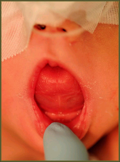
This is an 11-day-old baby who was presented by her parents to the Oral & Maxillofacial Surgery clinic at Children’s Hospital and Medical Center. Their chief complaint was that the baby’s tongue was progressively increasing in size, making it difficult for the mother to continue breastfeeding.
Sorry! you are incorrect
The location and the clinical description of “soft consistency” is characteristic of a ranula. The histology, however, is not supportive of a ranula.
Mucoceles and ranulas are clinical terms describing exophytic, fluid-filled, fluctuant nodules, typically of minor salivary gland origin and present mostly on the lower lip and the floor of the mouth (1-2). Over 90% of these lesions are cyst-like structures, or pseudocysts, and are mucous extravasation phenomena referred to as mucoceles. Some of these lesions are true cystic structures lined by epithelium and filled with mucus and are called mucus retention cysts or salivary duct cysts. These constitute a small percentage of all mucoceles (1-3). Ranulas, mucoceles of the floor of the mouth, constitute the other 5% and are divided into those above the mylohyoid muscle (majority) and below the mylohyoid muscle (also known as plunging ranulas or cervical ranulas) (3). Ranulas are of minor or major salivary gland origin and are mostly extravasation in type. The etiology of the extravasation mucoceles is usually sharp trauma cutting through the salivary gland duct and releasing the mucous in the extracellular tissue (1-2). Histologically, the extravasation-type mucocele consists of a cyst-like structure lined by granulation tissue and filled with mucoid material, foamy macrophages, and at times small clusters of neutrophils. The mucous retention cysts develop as a result of a duct blockage which can be caused by trauma, fibrosis, sialolith, or pressure from an overlying tumor (1). The extravasation mucoceles most commonly occur on the lower lip and very rarely on the upper lip. They may occur anywhere else in the oral cavity, including the buccal mucosa and floor of mouth (Ranula). The latter can be of minor salivary gland or submandibular or sublingual gland duct origin (2-3) and is more commonly seen in children and adolescents. It presents as a swelling with a bluish color if superficial, while deep mucoceles tend to take the color of the surrounding mucosa. Mucoceles tend to fluctuate in size. They are usually associated with a history of sharp lip or cheek biting, but can also be secondary to surgery in the area. [repetitive?] This is especially true with the anterior tongue mucoceles. Surgical excision with the associated minor salivary gland is the preferred treatment for the deep mucoceles; superficial mucoceles can self-heal within 2-3 weeks. Superficial mucoceles can also mimic vesiculobullous-type diseases because they look like vesicles (1), especially when presenting in multiples (rare, but described). They can recur if the source of trauma is not eliminated or if they are secondary to surgery. Simple (non-plunging) ranula is best treated by marsupialization into the floor of mouth (1-3). Plunging ranula requires complete excision via an extra-oral approach. The technical difficulties associated with the complete removal of this thin-walled lesion result in a relatively high recurrence rate.
Sorry! you are incorrect
The clinical description of “soft consistency” is consistent with a lipoma. The gross morphology (Fig 2) is suggestive of a lipoma. However, the histology is not supportive of a lipoma. Although rare in the mouth, this lesion has been described in the floor of mouth. Lipomas are also rare in children. Lipomas are benign neoplasms of adipose tissue origin (6-8). They are more commonly described in the trunk and extremities (8) and are rare in the oral cavity. Their overall incidence in the oral cavity is around 4.4% of all benign oral lesions (7). In a recent study from the Armed Forces Institute of Pathology (AFIP) of 125 benign lipomas in and around the mouth, the male to female ratio was approximately 3:1, which is not surprising given the study was extracted from a Military population. Other studies reveal an equal gender distribution (6). The AFIP study showed the mean age to be 52 with a range of 9-92 years (7). Only 4 of the 125 cases involved patients under 18 (8). In the mouth, the most common location for this neoplasm is the buccal mucosa followed by lips, submandibular area, tongue palate and less on the floor of mouth and vestibule (6-8). These findings are consistent with many other published reports. These lesions are slow-growing and can be present for many years. Lipomatous nodule of the buccal mucosa may represent herniation of the buccal fat pad. Lipomas usually present as a single, smooth surfaced, soft with doughy consistency, lobulated, painless, yellowish, sessile nodule (8). The overlying mucosa is usually thin and stretched with visible blood vessels. Because of its softness it can be mistaken for a cyst. Histologically, lipomas are variable benign histologies. Some have predominant lobules of mature adipocytes surrounded by a thin connective tissue capsule, while others have predominant spindle-cell component, myxoid, chondroid, connective tissue component. Some are intramuscular (8). Each has its own clinical behavior. Simple surgical excision is the treatment of choice for the simple, mature adipocyte component.
Sorry! you are incorrect
The most common teratoma of the mouth is the cystic teratoma known as dermoid cyst. They are rare under the age of 10. The histology is not supportive of a teratoma including dermoid cyst. Dermoid cysts of the oral cavity are rare and constitute around 1.6% of all dermoid cysts according to the original 1937 report by New and Erich (4). They are more common in the testis than in any other location, followed by the ovaries and the head and neck area (4-5). In the latter area, the floor of mouth is one of the more common areas of occurrence. This cyst is clinically classified into three types and is based on its relationship to the floor of mouth muscle, the geniohyoid and mylohyoid muscles (5). The more common presentation is above the geniohyoid and mylohyoid muscles, which are clinically visible (as is the case in this patient) in the floor of mouth as they push the tongue upward, leading to dysphagia, dyspnea and dysphonia. If the cyst is between the geniohyoid and the mylohyoid muscle or below the mylohyoid muscle, it creates the appearance of a double chin. The third type is where the cyst is displaced laterally into the submandibular area (4). Dermoid cysts of floor of the mouth are rarely described in children under the age of 10. The majority occur in patients between the ages of 10 and 30 for the floor-of-mouth cases and 15 and 40 for cases in the ovaries. Dermoid cysts above the geniohyoid muscle present as slowly enlarging large, round, raised and smooth-surfaced nodules. The nodules are usually painless unless infected, when pain can set in. Infected cysts can drain through either intraoral or extraoral fistulas. The size of the lesion determines its interference with eating, speaking and swallowing. This cyst is classified histologically into two types: cystic structures with a lumen filled with keratin and a connective tissue wall with skin adnexa and true cystic teratomas with all three germ layers tissues such as the brain, bone, muscle, respiratory and gastrointestinal tissues. The oral Dermoid cysts tend to be simple with skin adnexa in the wall. The true teratoma type cysts (more in the ovary) are prone to malignant transformation more so than those in the floor of mouth. Treatment of choice is surgical removal via intra- or extra-oral approach, depending on where the cyst is located.
Congratulations! You are correct
Lymphangioma is a benign congenital malformation of lymph vessels which is believed to be more of a hamartoma than a neoplasm. It constitutes 4% of all vascular tumors (9-10). Five types are described: lymphangioma circumscriptum (common in the tongue), cavernous lymphangioma (also common in the tongue), cystic hygroma (a large type of cavernous lymphangioma which is common in the head and neck area), acquired progressive lymphangioma, and lymphangiomatosis. These lesions can occur on the skin and mucosa and can be superficial (e.g. circumscriptum) or deep (e.g. cystic hygroma) (9). The head and neck area is the most common location for these lesions followed by the extremities and buttocks. In the oral cavity, the lymphangioma circumscriptum is superficial and presents in a “cobblestone” appearance on the dorsal tongue; it has a high recurrence rate. If it occurs on the lip it resembles angioedema. The lesion also occurs in the neck, deep into the tissue in a diffuse manner, which can be life threatening due to progressive airway compromise. This is called cystic hygroma. Lymphangioma, like hemangioma, is mainly a disease of children (about 90% develop in the first two years of life) (10); it also occurs in newborn babies, especially those of African descent (11). It is another major reason for macroglossia.
Cavernous lymphangioma is the diagnosis of this case; it usually occurs at infancy, more frequently in the head and neck areas and especially the tongue. It tends to be deep seated with intact and smooth mucosa which is different from the cobblestone appearance of the superficial lymphangioma. Surgery and laser surgery are the treatment of choice. Sclerosing agents have been described in treating large lymphangiomas successfully.
Treatment
Under general anesthesia and with an intra-oral approach, the lesion was completely excised (Fig 2). The lesion was soft and myxoid to palpation. The surgical site was closed with Vicryl suture around a 1.5cm Penrose drain. A four-week post-surgery visit revealed complete healing. The parents indicated that the patient began breastfeeding again.
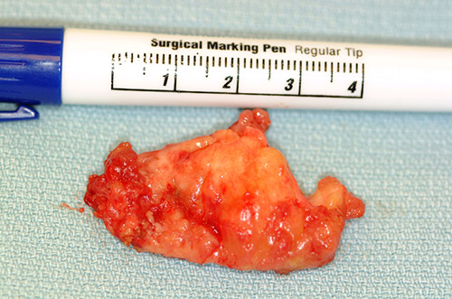
Figure 2. This is a gross specimen photograph of the excisional surgical specimen. Note the yellow color simulating fat. It is most likely the lesion surrounded by fat from the area.
References
- McGurk M. Management of the ranula. J Oral Maxillofac Surg. 2007 Jan;65(1):115-6.
- Chidzonga MM, Mahomva L. Ranula: experience with 83 cases in Zimbabwe. J Oral Maxillofac Surg. 2007 Jan;65(1):79-82.
- Loney WW Jr, Termini S, Sisto J. Plunging ranula formation as a complication of dental implant surgery: a case report. J Oral Maxillofac Surg. 2006 Aug;64(8):1204-8
- New, G. B., and Erich, J. B. Dermoid cysts of head and neck. Surg. Gynecol. Obstet. 65: 48, 1937
- Meyer, I. Dermoid cysts (dermoids) of the floor of the mouth. Oral Surg. Oral Med. Oral Pathol. 8: 1149, 1955
- Furlong MA. Fanburg-Smith JC, Childers ELB. Lipoma of the oral and maxillofacial region: Site and subclassification of 125 cases. Oral Surg Oral Med Oral Pathol Oral Radiol Endod. 2004 Oct;98(4):441-50
- Fregnani E.R, Pires FR, Falzoni R, Lopes MA and Vargas PA. Lipomas of the oral cavity: clinical findings, histological classification and proliferative activity of 46 cases. Int J Oral Maxillofac Surg 32 (2003), pp. 49–53.
- Fletcher C.D.M. Unni KK and Mertens F. Adipocytic tumors. In: Pathology and genetics: tumours of soft tissue and bone. World Health Organization classification of tumours, IARCPress, Lyon, France (2007), pp. 9–46
- Fletcher C.D.M. Diagnostic histopathology of tumors. Third edition, Churchill Livingstone Elsevier. Vascular tumors. pp 67-70.
- Neville B. Textbook of oral & maxillofacial pathology. Second edition (2002). Saunders, pp 475-476.
- Lo Muzio L, Mignogna MD et al. A rare case of fibrosarcoma of the jaws in a 4-year-old male. Oral Oncol. 1998; 34: 383-386
- Chigurupati, R, Aflatooni, A et al. Rhabdomyosarcoma of the head and neck in children, review of literature and addition of four cases. J Oral Oncology. 2002; 38: 508-515.
Sorry! you are incorrect
Fibrosarcoma
Fibrosarcoma is rare malignant neoplasm of fibrous connective tissue origin; only 10% of cases occur in the head and neck area. It generally occurs in patients between 20 and 40 years of age, but can occur in infants. It is slightly more common in males. It can occur in the buccal mucosa, maxillary sinus, palate, lips, or the periosteum of the mandible and maxilla. Sarcomas in general present as fleshly, polypoid, rapidly growing, ulcerative swellings that cause facial asymmetry. They can destroy bone. The clinical presentation and age of the patient in this case are consistent with fibrosarcoma; however, the histology is not supportive of it. Fibrosarcoma tends to present with pleomorphism and high mitotic activity (11).
Rhabdomyosarcoma
Rhabdomyosarcoma is the most common sarcoma in children. It is a malignant neoplasm of skeletal muscle origin. Three types are described: embryonal, alveolar and pleomorphic. The embryonal and alveolar types are most common in children, while the pleomorphic is most common in adults. All types of rhabdomyosarcoma present as exophytic, polypoid, ulcerated, and rapidly growing lesions. This condition may occur at birth, in children, in teenagers or in young adults; it is rare after the age of 45. The head and neck area is a common location for occurrence; such instances constitute 40% of all cases. The orbit is the most common location, followed by the nasal cavity, the oropharynx and the oral cavity. Within the oral cavity, the palate is the most common location. The clinical presentation of this case is suggestive of rhabdomyosarcoma, but the histology is not supportive of it (12).