Ulcerated Swelling in the Hard Palate
Can you make the correct diagnosis?
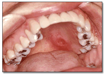
This is a 57-year-old Guatemalan female who complained of a palatal swelling that she first noticed eight months prior to the clinical consultation. The swelling was firm, ulcerated and asymptomatic.
Sorry! you are incorrect
Torus palatinus is a hyperplastic cortical bone swelling located in the midline of the hard palate and is hard on palpation. Patients usually give a history of its long term (many years’) presence. In one study it affected up to 38.5% of Asian females and 30% of white females (1). The etiology of this lesion is unknown, but heredity plays a role; some cases follow an autosomal dominant pattern of transmission.
Torus palatinus is a bony outgrowth at the midline of the hard palate covered by normal, sometimes thin, mucosa; at times, the mucosa undergoes ulceration. It can be smooth surfaced or multilobular, in which case it usually grows in four coalescing bony lobules. It is a slow-growing and painless lesion except if traumatized. The lesion occurs by age 30 and affects females twice as often as males. Larger lesions can interfere with speech, hygiene and dental prostheses. Radiographically, it is always radiopaque. Histologically, it is made up of dense viable bone with Haversian systems containing viable osteocytes.
Treatment is only indicated if it is symptomatic, interfering with function or creating difficulty with prostheses. These lesions are most easily removed by reflection of a mucoperiosteal flap and burring away with a hand piece. The clinical presentation, the consistency, the location and the histology are not supportive of torus palatinus.
Sorry! you are incorrect
Soft tissue swellings include both reactive and neoplastic (benign and malignant) lesions. The palate is a common location for soft tissue swellings, including those of fibrous connective tissue, peripheral nerve and muscle origin to name a few. One of the common fibrous connective tissue swellings is fibrous hyperplasia, denture or not denture related. Denture-related hyperplasia can be pedunculated and flat surfaced; it occurs in response to an ill-fitting maxillary denture and is treated with simple excision and denture reline or new denture formation. Peripheral nerve neoplasms such as neurofibromas and schwannomas can occur on both the anterior and posterior hard palate. Smooth and skeletal muscle neoplasms, especially rhabdomyosarcomas, can occur on the palate and are common in the head and neck area. They are especially common in the pediatric population. Lipomas may occur in the oral cavity, but rarely. They are especially rare on the palate (2). All of the lesions mentioned in this category can be ruled out based on the histology.
Congratulations! You are correct
Adenoid Cystic Carcinoma (ACC) Adenoid cystic carcinoma is a malignant neoplasm of epithelial salivary gland origin, predominantly of basal and myoepithelial cell origin. It occurs in minor and major salivary glands, including the parotid, submandibular, palatal, lacrimal, pharynx, laryngeal, skin, and breast glands among many other locations. It accounts for 4% of all salivary gland neoplasms and 7.5% of all epithelial salivary gland malignant neoplasms (5). There is no consensus in the literature on the most prevalent malignant salivary gland neoplasm of the oral cavity; some suggest adenoid cystic carcinoma to be the most common, while others show that mucoepidermoid carcinoma is the most common and yet a third group of reports suggest that polymorphous low grade adenocarcinoma is the most common (5, 7). It is not the intention of this author to favor any of these reports, but rather to state that adenoid cystic and mucoepidermoid carcinomas are more frequently diagnosed in our biopsy service than polymorphous low grade adenocarcinoma.
ACC occurs more commonly in middle-aged patients in the fourth to the sixth decade of life. However, it can occur in patients of all ages with slight predilection for occurrence in females. Although cases in the first decade and the tenth decade have been described, these extremes tend to be rare (5, 7-8). According to the AFIP’s records of their own diagnoses of ACC, 26.8% of cases occur in the parotid, 24% in the submandibular gland, 20.5% in the palate, 5% in the tongue, around 4% each in the lips and buccal mucosa and 1.2% in the sublingual gland (9).
Adenoid cystic carcinoma is a slow-growing neoplasm, persistent in growth and recurrence with a slow tendency for metastasis. It may present with local pain and facial nerve paralysis if affecting the parotid. Fixation to deeper structures and local invasion is common in the larger lesions. ACC of the palate may be covered with normal mucosa, making it indistinguishable from a benign salivary gland or soft tissue neoplasm. It may also be ulcerated, especially in larger lesions. It has a marked tendency for perineural invasion explaining the signs of pain or paresis (5-9).
Multiple genetic aberrations have been identified; one involves the loss of DNA copy number in chromosome 12q, and another involves deletion in 6q23 and 13q21 (6). Others show gain in chromosome 19 and deletion of chromosome 19q. Another study showed similarities in genetic abnormalities between adenoid cystic carcinoma and polymorphous low grade carcinoma involving chromosomes 12 and 6. These two malignant salivary gland neoplasms have other common clinical and histologic characteristics, making it difficult at times to differentiate between them.
Histologically, ACC is made up of infiltrative small, dark, uniform and basaloid cells with or without mitosis depending on the type. Three types have been described: cribriform pattern, solid pattern and tubular. Combinations of the three types are sometimes encountered in one neoplasm. Cribriform pattern is the most common type and is arranged in clusters and nests of epithelial cells with holes (spaces); thus, it is also known as a “Swiss cheese” pattern. The neoplastic cells are small, basaloid and uniform with little to no mitosis and no pleomorphism. It is clinically low-grade, behaving with multiple recurrences and a slow tendency for metastasis. The high-grade counterpart is also known as the solid type, where cells are arranged in nests with larger basaloid cells, pleomorphism and prevalent mitosis. Spaces within these clusters are usually scant or absent. This is a more aggressive type with a tendency for early metastasis. The tubular type is difficult to interpret since some believe is similar to the cribriform pattern in behavior. All three types have a tendency for perineural invasion (5-9).
Treatment
Complete surgical removal with wide clean margins of surrounding tissue is the treatment of choice. The patient elected a partial denture prosthesis with obturator. If there is histological evidence of perineural spread, nerves may be surgically followed to the skull base. Surgery combined with radiotherapy has been used, especially with recurrence or with surgical specimens involving the surgical margins. Radiotherapy alone has been used, but not effectively. Chemotherapy is used with metastasis. Prognosis is based on the clinical stage as well as the histology. Stages 1 and 2 have a much better 10-year survival rate than stages 3 and 4 (75% stages 1 and 2, 43% for stage 3 and 15% for stage 4). Histologically, the cribriform pattern has a better prognosis than the solid type. One study showed the 15-year survival rate for cribriform pattern cases to be 39%, compared with a 5% survival rate with the solid variant. The latter measured by tumor histologically made up of 30% solid ACC. These tumors tend to invade bone and metastasize to the lung. Because of the tendency for multiple recurrences and possible metastasis, long-term (20 years or more) follow-up is mandatory (5-9).
References
- Chohayeb AA, Volpe AR. Occurrence of torus palatinus and mandibularis among women of different ethnic groups. Am J Dent. 2001; 14: 278-280.
- Neville B, Damm D et al. Textbook of Oral & Maxillofacial Pathology. Saunders; 2002: second edition.
- Br Dent J. 2004; 196: 79-81.
- Sandmeier D, Bouzourene H. Necrotizing sialometaplasia: a potential diagnostic pitfall Histopathology. 2002;40:200-201.
- Bradley PJ. Adenoid cystic carcinoma of the head and neck: a review. Curr Opin Otolaryngol Head Neck Surg. 2004; 12: 127-132.
- El-Rifai W, Rutherford S et al. Novel DNA copy number losses in chromosome 12q12–q13 in adenoid cystic carcinoma. Neoplasia. 2001; 3: 173-178.
- Hyam DM, Veness MJ et al. Minor salivary gland carcinoma involving the oral cavity or oropharynx. Aust Dent J. 2004; 49:16-19.
- Ogawa Y, Kishino M et al. Adenoid cystic carcinoma associated with salivary duct cyst in the sublingual gland. J Oral Pathol Med. 2004; 33:311-313.
- Ellis G, Auclair P et al. Surgical pathology of the salivary glands. Saunders; 1991.
Sorry! you are incorrect
Necrotizing sialometaplasia is a spontaneous, self-healing, rapidly growing benign inflammatory lesion primarily affecting the minor salivary gland tissue. It usually heals within three months of occurrence. Clinically, however, it is an aggressive looking lesion. It is of unknown etiology, but transient local ischemia is suggested as a possible etiology. The latter is seen with recent dental injections to the area; the vasoconstrictor effect of dental injections is believed to induce local ischemia. The local infarction hypothesis is supported by the histology of necrotic acini with intact cell membrane which is seen in coagulative necrosis. It is more common in the fourth and fifth decade of life, and occurs more often in males (2:1 male: female ratio). The palate in general, and the junction of hard and soft palate in particular, are the most common locations of occurrence, accounting for about 75% of cases. The condition presents as single, bilateral (3) or multiple ulcers. The ulcers are deep and necrotic with a flat edge. They can reach more than three centimeters in size. The clinical symptoms, when present, include mild pain or parasthesia. The clinical presentation can be mistaken for a malignant neoplasm such as adenoid cystic carcinoma or surface squamous cell carcinoma. No treatment is recommended, but a biopsy is recommended to establish the baseline diagnosis. Most lesions heal within ten weeks after biopsy (4). The clinical presentation of this case is suggestive of necrotizing sialometaplasia; the histology, however, is not supportive. For more information on necrotizing sialometaplasia, please read the clinical case discussion section of the Summer 2004 e-PIE Newsletter.