February 2008: Exophytic red palatal gingival swelling tooth # 8
Can you make the correct diagnosis?
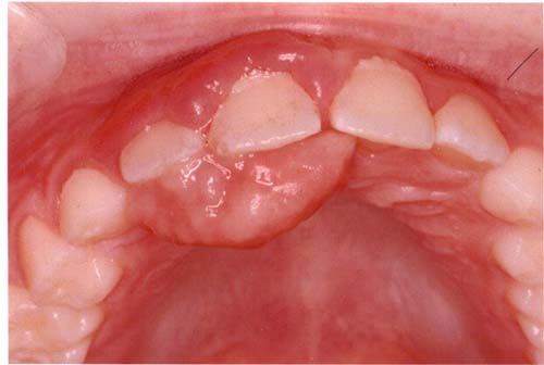
This is an otherwise healthy 8 year-old-boy who underwent a punch biopsy for a small gingival swelling which was removed in March 2007. The lesion recurred in September 2007 in a much larger and more rapidly growing form (Figure 1). This lesion measured 1 X 1.5 cm in size and was red and vascular in color compared to the previously excised lesion, which was 8 X 4 mm in size and pink in color. The current lesion originated between teeth #s 8 & 9. Both lesions were not painful but the recurrent lesion was large enough to interfere with speech and eating. Occlusal radiograph of the first lesion was negative for any bony involvement.
Sorry! you are incorrect
Pyogenic granuloma constitutes 85% of all reactive gingival swellings, representing a profuse mass of vascular granulation tissue (1). It can be induced by local irritants such as excessive plaque, sharp fillings and dental calculus; it sometimes forms in an extraction socket in response to an irritant left in the socket. It can occur anywhere in the oral cavity and skin, especially the tongue, lips, fingers and nail beds (1). In the mouth, it occurs most commonly in the gingiva, especially the maxillary buccal and interproximal gingiva (1-2). Occasionally, it may surround the tooth. It is usually highly vascular, fast-growing, exophytic, lobular, sessile, and ulcerated or covered by pseudomembrane. The color changes from red to pink when it starts to heal. It occurs at any age and sex with a slight predilection for young females; it affects 1% of pregnant females. Pyogenic granuloma is usually painless except during eating, when bleeding and pain is described (1). Histologically, it presents as a mass of loose and vascular granulation tissue, usually with ulcerated or eroded surface epithelium and many inflammatory cells. A range of treatment modalities are available, including excision with removal of the local irritant, laser surgery, or intralesional injection with absolute alcohol, steroids or botulinum toxin (2-3). Scaling and polishing prior to surgical removal helps shrink the lesion. The age, location and clinical presentation of this case are all supportive of pyogenic granuloma, but the histology is not.
You are correct!
Peripheral ossifying fibroma constitutes 10% of all reactive gingival swellings. It consists of a moderately cellular fibrous connective tissue mass with bony trabeculae and/or cementum-like hard tissue. It has been reported rarely on edentulous alveolar mucosa. It originates from the periodontal ligament or the periosteum. This lesion is most common in young patients between 1 and 19 years of age and has a predilection for females over males by a 3:2 ratio (7-9). It occurs exclusively on the gingiva, especially the anterior gingiva, with slight predilection to the maxilla and rare presentation in primary teeth (9). It is usually sessile and exophytic and often ulcerated; it presents as well-demarcated sessile nodules, which are firm or hard depending on the amount of ossification and calcifications (7-8). Peripheral ossifying fibroma is usually pink but can be focally red if ulcerated. Histologically, peripheral ossifying fibroma is made up of a moderately cellular mass of fibrous connective tissue with calcifications ranging from cementum-like material to calcified bony trabeculae with viable osteocytes. The surface epithelium overlying the mass is usually ulcerated. Deep surgical excision to include the periodontal ligament is the preferred treatment, though laser removal has been used effectively. Deep surgery may lead to a gingival defect, which would require gingival grafting, especially if it is located on the anterior buccal gingiva (8). There is a 16-20% recurrence rate (9).
References
- Fantasia JE, Damm DD. Red nodular lesion of tongue. Pyogenic granuloma.
Gen Dent. 2003 Mar-Apr; 51(2):190, 194. - Ichimiya M, Yoshikawa Y, Hamamoto Y, Muto M. Successful treatment of pyogenic granuloma with injection of absolute ethanol. J Dermatol. 2004 Apr; 31(4):342-4.
- Pham J, Yin S, Morgan M, Stucker F, Nathan CA. Botulinum toxin: helpful adjunct to early resolution of laryngeal granulomas. J Laryngol Otol. 2004 Oct; 118(10):781-5.
- Flaitz CM, Peripheral giant cell granuloma: a potentially aggressive lesion in children. Pediatr Dent. 2000 May-Jun; 22(3):232-3.
- Chaparro-Avendano AV, Berini-Aytes L, Gay-Escoda C. Peripheral giant cell granuloma. A report of five cases and review of the literature. Med Oral Patol Oral Cir Bucal. 2005 Jan-Feb; 10(1):53-7; 48-52.
- Neville BW, Damm DD, Allen CM, Bouquot JE. Peripheral giant cell granuloma. In: Oral and Maxillofacial Pathology, 2nd edition. Philadelphia: W.B. Saunders, 2002. p. 449-451.
- Hanemann JA, Pereira AA, Ribeiro Junior NV, Oliveira DT. Peripheral ossifying fibroma in a child: report of case. J Clin Pediatr Dent. 2003 Spring; 27(3):283-5.
- Walters JD, Will JK, Hatfield RD, Cacchillo DA, Raabe DA. Excision and repair of the peripheral ossifying fibroma: a report of 3 cases. J Periodontol. 2001 Jul; 72(7):939-44.
- Cuisia ZE, Brannon RB. Peripheral ossifying fibroma–a clinical evaluation of 134 pediatric cases. Pediatr Dent. 2001 May-Jun;23(3):245-8
- Bundgaard T, S Bentzen, et al. Histopathologic, stereologic, Epidemiologic, and clinical parameters in the prognostic evaluation of squamous cell carcinoma of the oral cavity. Head & Neck. 1996. 18:142-152.
- Barasch A, Morse DE. Smoking, gender, and age as risk factors for site-specific intraoral squamous cell carcinoma. Cancer 1994. 73:509-513.
- Syrjanen SM, Syrjanen KJ. Human papillomavirus (HPV) DNA sequences in oral precancerous lesions and squamous cell carcinoma demonstrated by in situ hybridization. 1988. J Oral Pathol. 17:273.
- Holmstrup P, JJ Thorn. Malignant development of lichen planus-affected oral mucosa. J Oral Pathol. 1988. 17:219-25.
- Chigurupati R, Alfatooni A, Myall RW, Hawkins D, Oda D. Orofacial rhabdomyosarcoma in neonates and young children: a review of literature and management of four cases. Oral Oncol. 2002 Jul;38(5):508-15.
- Uren A, Toretsky JA. Pediatric malignancies provide unique cancer therapy targets. Curr Opin Pediatr. 2005 Feb;17(1):14-9.
- Grohn ML, Borzi P, Mackay A, Suppiah R. Management of extensive congenital fibrosarcoma with preoperative chemotherapy. ANZ J Surg. 2004 Oct;74(10):919-21
Sorry! You are incorrect
Peripheral giant cell granuloma constitutes less than 5% of all reactive gingival swellings, and consists of a hyperplastic mass of vascular granulation tissue with many osteoclast-like multinucleated giant cells. It presents as a lobular, purplish-blue exophytic nodule exclusively on the gingiva, both edentulous and dentate, and usually anterior to the molars (4-5). It originates from either the periodontal ligament or the periosteum. It occurs across a wide age range, especially in children, young adults, and females (2:1 female to male ratio) (4-6). It presents as either sessile or pedunculated and smooth surfaced or lobular; though usually painless, it can occasionally be ulcerated, painful and accompanied by bleeding (4-6). Like pyogenic granuloma, it is usually present either on the buccal or lingual gingiva or between teeth, but it can occasionally surround the teeth (4-6) and act aggressively by displacing teeth much like a sarcoma (4). It can also resorb the underlying bone in a smooth and concave “saucer-like” manner. Complete excision including curettage of underlying bone is the preferred treatment. It has a good prognosis with recurrence rate of approximately 10% (6). The age, location and clinical presentation of this case are all supportive of peripheral giant cell granuloma, but the histology is not.
Sorry! You are incorrect.
Squamous cell carcinoma in a child of this age is extremely rare. In addition, SCC of the gingiva is uncommon. Oral squamous cell carcinoma (OSCC) of the mouth is a highly aggressive neoplasm that currently ranks as the fifth most common malignant neoplasm worldwide and accounts for an estimated 90% of oral malignancies (10). Oral SCC occurs predominantly in males over the age of 40 years, with an observed male-to-female ratio of 2:1 generally and 1.4:1 in the USA (10-11). Excluding the outer lip, the most common sites (in decreasing order) are ventral and lateral surfaces of tongue (25-50%), floor of mouth (15%), gingiva (12%) and palate (9%). The buccal mucosa and retromolar pad areas (3%) have a relatively low incidence of occurrence (12) unless the patient is a chronic smokeless tobacco user. Oral SCC varies in presentation from deceptively innocent-looking to obviously malignant. It may present as a non-healing ulcer, or as red, white or mixed red-and-white lesions. Characteristic signs of oral SCC are non-healing ulcer, ulcer with rolled borders, fungation, fixation and induration. Rarely, OSCC may present as unexplained asymptomatic lateral neck lymphadenopathy (10-12). Oral SCC is most commonly associated with chemically induced mutagenesis, specifically tobacco and alcohol use and others such as long-standing erosive lichen planus (13). The age, the location and the histology in this case are not supportive of SCC.
Sorry! You are incorrect.
Rhabdomyosarcoma, fibrosarcoma and other less aggressive mesenchymal (infantile myofibromatosis, aggressive fibromatosis) types of lesions should be considered when a lesion recurs in such a short period of time and especially in a child.
Rhabdomyosarcoma is a malignant neoplasm of skeletal muscle origin accounting for 4-8% of all malignant diseases in children less than 15 years of age. It has been reported that 35% of the head and neck rhabdomyosarcomas in children are misdiagnosed (14-15). It primarily occurs in the first decade of life with a peak incidence between 2 and 6 years, the majority of patients being under 12 years of age (14). It is slightly more common in males. The orbit is the most common location followed by the nasal cavity, oropharynx and the oral cavity. Rhabdomyosarcomas of the oral cavity account for 10-12% of the head and neck cases (15). The tongue, palate and cheek are the most common locations in the mouth (14). The clinical appearance ranges from a small cutaneous nodule to an extensive mucosal outgrowth. It may present as a painless, yet occasionally painful, facial swelling. The presenting clinical features are often non-diagnostic. They often mimic conditions like infection. Trismus, paresthesia, facial palsy, and aural or nasal discharges have also been described. The histology in this case is not supportive of a sarcoma.
Infantile Fibrosarcoma is a rare malignant neoplasm of fibroblast origin. An adult counterpart exists and is more aggressive, with a 40% five-year survival rate compared to the infantile five-year survival rate of 80% (16). Like rhabdomyosarcomas, they can be congenital and usually develop within the first two years of life. They are slightly more common in males. Only 10% occur in the head and neck area, including the oral cavity (16). They are more common on the distal portion of the extremities. In the oral cavity, the buccal mucosa, palate, lips and periosteum of the mandible and maxilla are the most common locations of occurrence. Fibrosarcomas present as fleshly, polypoid, rapidly growing, ulcerated lesions. They can cause asymmetry, tooth displacement, and bone and tooth resorption. The histology in this case is not supportive of a sarcoma.