Small flat dark brown lesion right anterior floor of mouth
Dolphine Oda, BDS, MSc
doda@u.washington.edu
Contributed by: Dr. Stephen Knoff
Kirkland Oral Surgery, Kirkland, WA
Case Summary and Diagnostic Information
This is a 53-year-old white female referred presenting for the evaluation of a 4 x 3 mm flat pigmented lesion on the floor of mouth.
Diagnostic Information Available
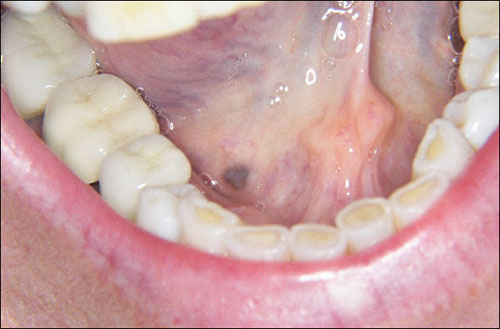
This is a 53-year-old white female referred by her dentist for the evaluation of a 4 x 3 mm flat, well-demarcated pigmented lesion on the right anterior floor of mouth (Figure 1). The lesion was of unknown duration and was asymptomatic. The patient stated, “I had no idea it was there until my dentist noticed it.” Clinically, the lesion was brown to blue/black in color. Her past medical history is significant for a hysterectomy 7 years previously. She has no history of tobacco use. The patient has no current medical problems, no medications and no allergies.
Figure 1. Clinical photograph of the lesion taken at the first visit. Note the flat, well-demarcated, dark brown lesion on the right anterior floor of mouth.
This patient’s past medical history is significant for a hysterectomy 7 years previously. She has no history of tobacco use and drinks occasionally.
This flat, well-demarcated, variably pigmented lesion on the right anterior floor of mouth was present for an unknown period of time and was otherwise asymptomatic. The variation in the color was of concern to the dentist, who therefore recommended a biopsy.
Figure 1. Clinical photograph of the lesion taken at the first visit. Note the flat, well-demarcated, dark brown lesion on the right anterior floor of mouth.
Treatment
Under local anesthesia, the lesion was completely excised and closed primarily. The results of the excisional biopsy were such that no further treatment was deemed necessary. The patient healed with no complications.
Excisional Biopsy
Histologic examination reveals a piece of soft tissue composed of keratinized surface epithelium with underlying fibrous connective tissue containing foreign material (Figures 2 & 3) This material is in the form of small aggregates of granular brownish black particles deposited on delicate collagen fibers especially around small blood vessels and the basement membrane (Figure 2). Some of this material is deposited on delicate collagen fibers in the superficial lamina propria (Figure 3).
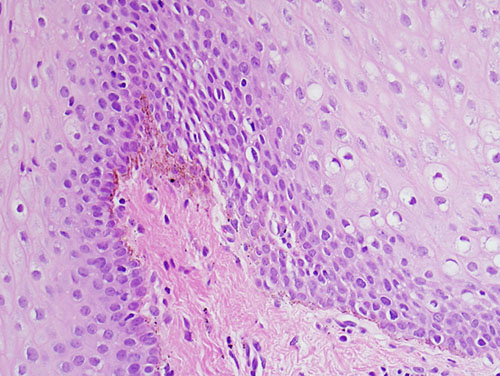
Figure 2. High power (x200) histology shows H & E stained section with surface epithelium and underlying fibrous connective tissue containing brownish-black granular material deposited on delicate collagen fibers of the basement membrane.
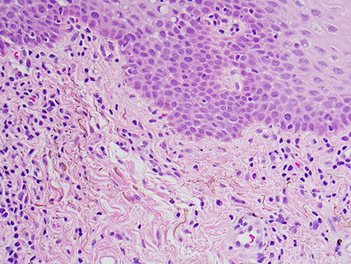
Figure 3. High power (x200) histology shows H & E stained section demonstrating the granular brownish black material deposited on delicate collagen fibers in the superficial lamina propria.
After you have finished reviewing the available diagnostic information