Return to Case of the Month Archives
Dome shaped, smooth-surfaced swelling, floor of mouth
Dolphine Oda, BDS, MSc
doda@u.washington.edu
Contributed by
Dr. Craig Neal
Neal Oral Surgery, Seattle, WA
Case Summary and Diagnostic Information
This is a 67-year-old male with a smooth-surfaced, dome-shaped and pink- to grayish-blue swelling located off the midline in the left side of the floor of mouth.
Diagnostic Information Available
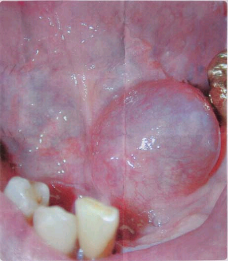
This is a 67-year-old male with a smooth-surfaced, dome-shaped and pink- to grayish-blue swelling located off the midline in the left side of the floor of mouth (Fig 1). The swelling is about 2 cm in diameter and has been present for an unknown period of time.
Figure 1. This photograph was taken at the primary clinical presentation. Ranula, floor of mouth presenting as dome-shaped, smooth surfaced, bluish grey swelling which is clinically an ideal presentation for this lesion in terms of color, shape and location.
This patient’s past medical history includes tonsillectomy and adenoidectomy at age 9, appendectomy at age 19, and several afflictions during adulthood including stomach ulcers and other gastrointestinal problems, breathing difficulties, stiff hip and right leg pain. He smokes one pack of cigarettes per week and drinks two bottles of wine per week. He also reports hypersensitivity to penicillin.
This smooth-surfaced, dome-shaped and pink- to grayish-blue swelling is described to be fluctuant. It is not painful and is present off the midline in the floor of mouth. The swelling is about 2 cm in diameter. The CT scan images showed no evidence of a sialolith in the sublingual or submandibular salivary gland ducts.
Histologic examination revealed a cyst-like structure surrounded by minor salivary gland lobules. The latter shows evidence of connective tissue fibrosis, chronic inflammation and ductal ectasia (Fig 3). The cyst-like structure occupies the bulk of the specimen and is large and tortuous. It is made up of a rim of granulation tissue surrounding a lumen that focally contains mucoid material and foamy macrophages (Fig 2).
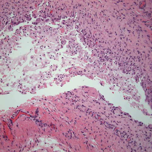
Figure 2. Low power (x100) histology shows a cyst-like structure lined by granulation tissue and filled with mucoid material with sheets of foamy macrophages.
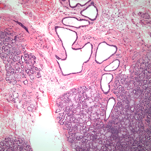
Figure 3. High power (x200) histology shows the lumen of the cyst-like structure filled with mucoid material and foamy macrophages.
After you have finished reviewing the available diagnostic information