Single grayish-brown swelling between teeth #s 5 & 6
Dolphine Oda, BDS, MSc
doda@u.washington.edu
Contributed by: Dr. Stephen Knoff
Kirkland Oral Surgery, Kirkland, WA
Case Summary and Diagnostic Information
This is a 14-year-old white male who presents with an exophytic, grayish-brown swelling between teeth #s 5 & 6.
Diagnostic Information Available
This 14-year-old white male was referred to the oral surgeon in October 2008 for surgical exposure of impacted teeth #s 6 and 11 and placement of brackets with gold chains to facilitate orthodontic eruption. The maxillary incisor teeth showed severe resorption. Surgical exposure with bracket placement was accomplished in January 2009. In May 2010 the bracket with gold chain on tooth #6 had come off and the patient was referred back for replacement (Figure 1). The alveolar mucosa overlying tooth #6 was swollen and inflamed; this tissue was debrided and a new bracket with chain was placed. The patient was referred again in June 2011 to evaluate an exophytic, grayish-brown gingival swelling between teeth #s 5 & 6 (Figure 2). The area was asymptomatic and the lesion was described to be 7 x 10 mm in size.
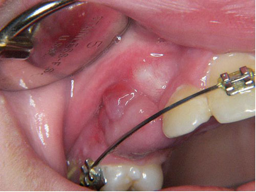
Figure 1. This photograph was taken in May 2010 where a small pinkish-red swelling is identified in the area of unerupted tooth #6.
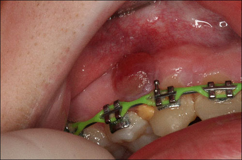
Figure 2. This photograph was taken in June 2011, one year after the small swelling in figure 1 was debrided. This is the recurrent lesion. The swelling on the buccal gingiva between teeth #s 5 & 6. It is exophytic and has a grayish brown color.
The patient’s past medical history is significant for hypersensitivity to sulfa drugs and penicillin. The patient is a non-smoker.
As described in the patient’s present history, this lesion was present a year prior to the current lesion and was in the area of unerupted tooth #6 (Figure 1). It was debrided but it slowly recurred over a one-year period. The second lesion was a grayish-brown and asymptomatic swelling (Figure 2) and was described as 7 x 10 mm in size.
Figure 1. This photograph was taken in May 2010 where a small pinkish-red swelling is identified in the area of unerupted tooth #6.
Figure 2. This photograph was taken in June 2011, one year after the small swelling in figure 1 was debrided. This is the recurrent lesion. The swelling on the buccal gingiva between teeth #s 5 & 6. It is exophytic and has a grayish brown color.
Treatment
Under local anesthesia, the lesion was excised sharply and the roots of teeth #s 5 and 6 were scaled to ensure complete removal of any local irritants. It was allowed to heal by secondary intention.
Excisional Biopsy
Histologic examination reveals a hemisected piece of soft tissue composed of keratinized and ulcerated surface epithelium with an underlying granulation tissue mass with many giant cells (Figure 3). The surface also shows a large area of ulceration covered by fibrin and neutrophils. The underlying granulation tissue mass is loose and vascular and contains numerous giant cells of variable shapes and sizes (Figure 4). The giant cells are haphazardly arranged. The granulation tissue mass contains clusters of extravasated erythrocytes as well as clusters of hemosiderin pigment (Figure 5). This mass and the surrounding connective tissue are infiltrated by lymphocytes, plasma cells, and neutrophils.
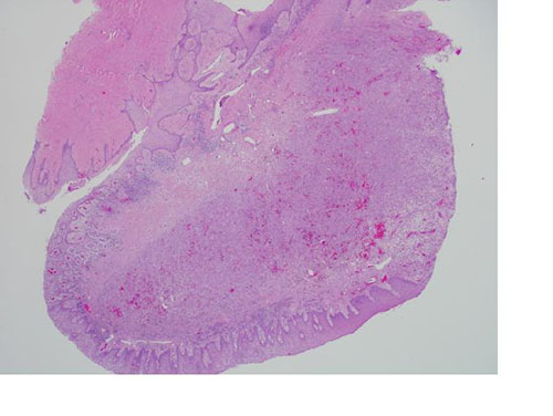
Figure 3. Low power (x40) H & E stained histology shows a mass of vascular granulation tissue with many multinucleated giant cells and ulcerated surface epithelium.
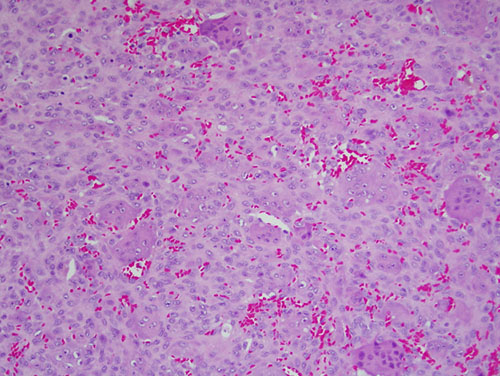
Figure 4. Higher power (x100) H & E stained histology shows sheets of multinucleated giant cells suspended on a background of vascular granulation tissue.
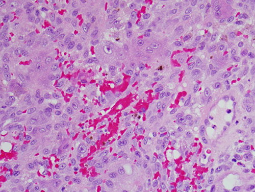
Figure 5. High power (x200) H & E stained histology shows small clusters of hemosiderin pigment and clusters of extravasated erythrocytes in a background of granulation tissue and giant cells.
After you have finished reviewing the available diagnostic information