Generalized Ulcerative Oral Mucosa
Can you make the correct diagnosis?
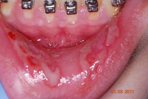
This is a 14-year-old white male who was referred to the Department of Oral Medicine, UW SOD with a chief complaint of not being able to eat or drink because of generalized oral ulcers and sloughing epithelium that had been present for eight weeks.
Sorry! you are incorrect
Acute and generalized oral ulcers in a 14-year-old would make one think a viral etiology as the cause. In this case, primary herpetic gingivostomatitis would be the most likely viral disease. Lack of fever even at the start of the disease and the eight-week or more duration speaks against herpetic gingivostomatitis given that the patient was otherwise healthy. The histology was not supportive of this condition.
Primary Herpes simplex virus1 (HSV1) is the most common acute viral infection of the oral mucosa, affecting both children and young adults. More than 80% of cases are sub clinical, in that the person exposed to the virus will not show evidence of disease with the exception of mild common cold-like symptoms. However, once an individual is infected, the virus will remain in the body for life. The body, however, develops antibodies to the virus within the first two days to three weeks of exposure and it is usually IgM and IgG. Medications such as acyclovir treat the symptoms of the disease and may weaken the virus and delay recurrence, but acyclovir and other drugs in this family will not eradicate the virus. HSV1 usually affects children congested developing countries and adolescent and young adults in developed countries such as the US. In the US 60% and the adult population is infected with HSV1 compared to 90% of the adult population developing countries. This virus also causes genital herpes and is called Herpes simplex 2 (HSV2). Herpes simplex 1 can also infect the genital area while HSV2 can also infect the oral cavity. It is very difficult to tell the difference between the two clinically, but DNA sequencing can be more specific. They both present in the form of small, painful vesicles that rupture within a short period of time. The fluid released by the ruptured vesicles releases virions that are live viruses capable of infecting others via contact (i.e. kissing, sharing food or drink). A person can autoinoculate by touching the open vesicle and then touching other areas before washing their hands. It is very important to wear gloves before touching a lesion believed to be of viral etiology, and to wash hands afterwards. The small percentage of the patients that manifest the disease present with small painful blisters, fever up to 103° F, drooling, headaches, pain upon swallowing, cervical lymphadenopathy, and extremely inflamed mucosa or skin. The numerous small vesicles are more common on the palate and dorsal tongue, but can be present anywhere. They rupture within one or two days to form shallow, ragged, painful, small ulcers with uniform size. Some coalesce to form irregular larger superficial ulcers. They heal in 10-14 days, after which the virus travels along the nerve to remain latent in the ganglion supplying the area. HSV-1 usually travels along the trigeminal nerve and harbors the trigeminal ganglion, while HSV-2 harbors the lumbosacral ganglion. Patients can also develop complications such as pneumonia and meningitis. The vesicles break, releasing a yellow fluid replete with live viruses that can be cultured or smeared on a glass slide for cytological evaluation. The cytology typically shows epithelial cells with ballooning degeneration, intranuclear inclusion bodies (Lipschutz bodies), and epithelial cells with multiple nuclei. These changes can also be observed if a biopsy is taken. In mild cases, supportive treatments such as rest, fluids, and soft diets, along with either topical tetracycline rinses multiple times a day or systemic antibiotics (if secondarily infected) are used to expedite healing. In severe cases, systemic Acyclovir can be very helpful, especially if used in the early stages of the disease. In HIV patients and other immune compromised patients, IV acyclovir is recommended.
Sorry! you are incorrect
The severity and generalized nature of the ulcers, the duration of longer than eight weeks, and the lack of fever would raise the question of whether or not this is SJS and therefore should be included high on the differential diagnosis list. The histology of SJS is usually non-specific. For that reason, it was difficult to rule it out in this first biopsy, but it was ruled out in the second biopsy.
Any drug, including over the counter drugs, can cause Stevens-Johnson Syndrome (SJS) and Toxic Epidermal Necrolysis (TEN), both of which represent a severe form of erythema multiforme. SJS is a skin and mucous membrane disease that can cause rashes, bullae, ulcers, and epithelial sloughing (skin peeling which is more common in TEN). It is an IgE immune-mediated hypersensitivity reaction most frequently caused by drug intake, especially by sulfa drugs such as bacterim taken for urinary tract infections. SJS may also be a sequel to viral or mycoplasma infections, and occasionally to an underlying malignant neoplasm or a gastrointestinal disease. Skin lesions are usually the first symptoms to appear, followed by blistering and ulceration of the mucous membrane, although the reverse is also possible. They present with a rash on the extremities, progressing to the back, buttocks, chest, and other skin areas. The mucous membrane includes the mouth, eyes (SJS may lead to blindness), genitalia, the gastrointestinal tract, and the respiratory tract. The latter two are serious, especially if they lead to esophageal stricture and respiratory failure because of severe epithelial sloughing. In the oral cavity, SJS is more related to drug reactions, especially to sulfa drugs. It presents as acute or chronic types with varying degrees of severity. It may be life-threatening, especially in children. The antibodies target the epithelium and the superficial blood vessels, leading to epithelial ulceration, sloughing, and vascular leakage. Clinically, it is most common in young males (2:1 M:F ratio) between the ages of 20 to 40. It may also affect children. Diagnosis is often made on the basis of the clinical presentation. Neither the hematoxyline and eosine histology nor the immunofluoresence stains are specific. Treatment includes the withdrawal of the causing drug (if determined), fluids, supportive care, systemic steroids, and immune suppressants, dependent on severity. The bullae and ulcers may continue to appear even after the causative agent is discontinued. It takes six to eight weeks for the epithelial lesions to heal. The prognosis is determined clinically on a scoring system known as SCORTEN (grades 1-5): the higher the number, the worse the prognosis. For example, the mortality rate of grade 5 is over 90%.
Sorry! you are incorrect
This patient has had orthodontic treatment in the past with no reaction to the metal braces or metal wire. Two weeks prior to the ulcers, he was placed on new braces and flexible (nickel containing) wire. It would be natural to include hypersensitivity reaction on the differential diagnosis given that nickel is the most common of the metals causing hypersensitivity reaction. The nickel containing orthodontic wire was removed by the orthodontist soon after the ulcers began but the ulcers continued to appear and progress. Dr. Martin twice performed standard dermal patch testing using allergens for nickel and chromium according to ICDRG (International Contact Dermatitis Research Group) standards for type IV sensitivity. In both cases the reactions to both allergens was negative. The histology of the second biopsy was not supportive of a hypersensitivity reaction.
Hypersensitivity, also known as allergy, is a reaction to drugs, food or other substances, mediated by immunological reactions. There are several types of hypersensitivity reactions including anaphylactic or immediate type hypersensitivity (type I), cytotoxic (type II), immune complex disease (type III) and cell mediated hypersensitivity or delayed type (type IV). Immediate hypersensitivity (anaphylactic type) is mediated by memory IgE antibodies that drill holes in mast cell membrane, leading to the release of vasoactive amines causing vasodilatation and smooth muscle contraction thus edema and difficulty breathing. Type II (cytotoxic type) involves antibodies such as IgG and IgM binding to a surface antigen causing damage to the “surface” of the cells, predisposing them to phagocytosis or opsonization and cell lysis; examples include hemolytic anemia and erythroblastosis fetalis. Type III (immune complex mediated) is an antigen and antibody reaction that activates serum mediators, especially the complement system. The antigen-antibody complex formed in the blood can be removed by the phagocyte system if large enough or can start a very serious and life-threatening process of depositing the complexes in a variety of tissues such as blood vessels, kidneys and joints. The deposition of the complexes will generate an inflammatory process; examples of type III includes arthus reaction, serum sickness, systemic lupus erythematosus and acute glomerulonephritis among many. Type IV (cell mediated or delayed type) hypersensitivity is applied in this case. It is a result of sensitized T lymphocytes to a protein (most commonly surface but can be otherwise) such as keratin, thus leading to contact dermatitis or stomatitis. Surface contact hypersensitivity is the common example but tuberculosis and organ transplant rejection are among other examples.
Congratulations! You are correct
Although pemphigus vulgaris is much more common in adult female patients (2:1), it can occur, but rarely, in children. This raises the question of paraneoplastic pemphigus as a possible diagnosis. The typical clinical presentation of paraneoplastic pemphigus involves mucous membrane and skin lesions similar to Stevens-Johnson Syndrome (SJS), which was not the case in this patient. It is, however, important to mention that paraneoplastic pemphigus cases confined to the oral cavity have been described in patients with Hodgkin’s lymphoma and Castleman’s disease. The patient’s general health did not warrant a workup for an underlying malignancy, but he will be followed for any new signs and symptoms.
Pemphigus vulgaris is the most common lesion of the pemphigus family. There are various other pemphigus subtypes including vegetans, foliaceus erythematosus, and paraneoplastic. This condition is less common in the mouth than in the lichen planus or mucous membrane pemphigoid. It is an autoimmune disease that causes desquamation of the oral mucosa and skin. The antibody targets intercellular desmosomal adhesion molecules called desmogleins, which results in the breakdown of the middle part of the spinous layer, leaving the basement membrane intact. This is histologically referred to as acantholysis and suprabasilar separation.
Pemphigus vulgaris is a serious and life-threatening disease and can kill the patient if not treated. It has a mortality rate of 60-90% in non-treated patients, compared to 5% in treated patients. Complications from steroid treatment and infection can be fatal. Patients with pemphigus vulgaris also develop circulating antibodies; the antibody titer in this disease may be related to the severity of the local disease. Drugs such as penicillamine can induce PV, while neoplasms such as leukemia, Hodgkin’s lymphoma, and Castleman’s disease can induce paraneoplastic pemphigus. Several proteins are targeted in paraneoplastic pemphigus.
This disease has a high prevalence in patients of Jewish descent, as well as patients of Mediterranean descent (such as Greeks and Italians), with females being affected more commonly than males (2:1 F:M ratio). It occurs more often in patients of ages 40 to 60, and rarely occurs in children, though this has been described. Oral lesions are usually the first to occur, as is the case in this patient. In the mouth, the soft palate and buccal mucosa are common sites for PV, though it can also affect the gingiva and lateral tongue, mainly because of trauma from brushing and chewing on the lateral tongue. The erosions and sloughing epithelium can be painful especially with hot drinks, alcoholic beverages, and spicy or acidic food. These patients have difficulty brushing their teeth. PV affects other mucosae including the nasopharynx, anogenital, and esophagus. Skin lesions frequently present with vesicles and bullae that easily rupture, forming ulcers. The Nikolsky sign is positive in patients with PV.
This condition presents with supra-basilar clefting, leading to splitting of the epithelium above the basal cell layer, as well as blister formation. This is the result of acantholysis in the middle spinous layer epithelial cells, releasing cells within the cleft known as Tzanck cells. Immunoflourescent (IMF) studies, both direct and indirect, can be useful in reaching a specific diagnosis. By direct IMF, the spinous layer cells are positive with IgG and C3 forming a fishnet pattern around the epithelial cells. Treatment includes systemic and local steroids, as well as immune suppressant-type medications, dependent on the severity of the disease.
References
- Usatine RP, Tinitigan R. Nongenital herpes simplex virus. Am Fam Physician. 2010 Nov 1;82(9):1075-82.
- Horowitz R, Aierstuck S, Williams EA, Melby B. Herpes simplex virus infection in a university health population: clinical manifestations, epidemiology, and implications. J Am Coll Health. 2010 Sep-Oct;59(2):69-74.
- Tovaru S, Parlatescu I, Tovaru M, Cionca L. Primary herpetic gingivostomatitis in children and adults. Quintessence Int. 2009 Feb;40(2):119-24.
- Majorana A, Bardellini E, Flocchini P, Amadori F, Conti G, Campus G. Oral mucosal lesions in children from 0 to 12 years old: ten years’ experience. Oral Surg Oral Med Oral Pathol Oral Radiol Endod. 2010 Jul;110(1):e13-8. Epub 2010 May 10.
- Osterne RL, Matos Brito RG, Pacheco IA, Alves AP, Sousa FB. Management of erythema multiforme associated with recurrent herpes infection: a case report. J Can Dent Assoc. 2009 Oct;75(8):597-601.
- Latsch K, Girschick HJ, Abele-Horn M. Stevens-Johnson syndrome without skin lesions. J Med Microbiol. 2007 Dec;56(Pt 12):1696-9.
- Jawetz RE, Elkin A, Michael L, Jawetz SA, Shin HT. Erythema multiforme limited to the oral mucosa in a teenager on oral contraceptive therapy. J Pediatr Adolesc Gynecol. 2007 Oct;20(5):309-13.
- Pazzini CA, Pereira LJ, Marques LS, Generoso R, de Oliveira G Jr. Allergy to nickel in orthodontic patients: clinical and histopathologic evaluation. Gen Dent. 2010 Jan-Feb;58(1):58-61.
- Ehrnrooth M, Kerosuo H. Face and neck dermatitis from a stainless steel orthodontic appliance. Angle Orthod. 2009 Nov;79(6):1194-6.
- Kolokitha OE, Chatzistavrou E. Allergic reactions to nickel-containing orthodontic appliances: clinical signs and treatment alternatives. World J Orthod. 2008 Winter;9(4):399-406.
- Lara-Corrales I, Pope E. Autoimmune blistering diseases in children. Hautarzt. 2009 Mar;60(3):208-16. Semin Cutan Med Surg. 2010 Jun;29(2):85-91.
- Cervini AB, Tosi V, Kim SH, Bocian M, Chantada G, Nousari C, Carballo OG, Pierini AM. [Paraneoplastic pemphigus or paraneoplastic autoimmune multiorgan syndrome. Report of 2 cases in children and a review of the literature]. Actas Dermosifiliogr. 2010 Dec;101(10):879-86.
- El Fekih N, Kharfi M, Karaa A, Kamoun MR. Complete recovery from juvenile pemphigus vulgaris. [Indian J Dermatol Venereol Leprol. 2008 Nov-Dec;74(6):654-5.