Multilocular Radiolucency; Left Posterior Mandible
Can you make the correct diagnosis?
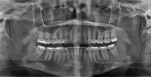
This is a 44-year-old old female who was seen by her general dentist on July 16, 2010 and was referred because of a radiolucent lesion noted on the panoramic radiograph.
Sorry! you are incorrect
The corticated multilocular radiolucency, the location and the age of the patient all support a diagnosis of ameloblastoma; therefore, it should be placed highest on the differential diagnosis. However, the mild expansion is not typical of ameloblastoma. Ameloblastomas tend to be clearly expansile. The histology was also not supportive of ameloblastoma.
Ameloblastoma is one of the most common benign neoplasms of odontogenic origin. It accounts for 11% of all odontogenic neoplasms/hamartomas (1-5). It is a slow-growing, persistent, and locally aggressive neoplasm of odontogenic epithelial origin. It affects a wide range of age distribution but is mostly a disease of adults, at an average age of 33, with equal sex distribution. Reports from Africa and India show a male predilection; it also has a predilection for occurrence in black patients (3-5). About 85% of ameloblastomas occur in the posterior mandible. Most of these occur in the molar-ramus area, while some occur in the anterior mandible. Three types of ameloblastoma are described. The most common is the multilocular solid type, constituting 86% of all ameloblastomas, which is characteristically expansile, radiolucent and multilocular. The unicystic type is the next most common type, constituting 13% of all ameloblastomas. It is radiographically unilocular and is associated with the crown of an impacted tooth in 90% of cases (1-4). Unicystic ameloblastoma occurs in patients around 14-20 years of age. The third type is peripheral ameloblastoma which can affect a wide age range and occurs mostly on the gingiva, simulating a reactive gingival swelling such as a peripheral ossifying fibroma or pyogenic granuloma. Ameloblastoma, if not treated, can reach very large sizes, causing facial disfigurement. It loosens, displaces and resorbs adjacent teeth. Ameloblastomas are usually not painful unless infected, in which case they can be mildly painful. Parasthesia and anesthesia are extremely rare, unless the lesion is very large in size. Also, ameloblastoma tends to expand rather than perforate the cortical bone; if the latter occurs with extension into the adjacent soft tissue, it has a higher tendency for recurrence and therefore a worse prognosis than cases in which the ameloblastoma is completely encased by bone (1-5). The solid type is treated with en bloc or resection with clean margins. Curettage is the treatment of choice for the unicystic type.
Sorry! you are incorrect
It is typical for CGCG to present as a multilocular radiolucency anterior to the mandibular first molar. CGCG should therefore be seriously considered on the differential diagnosis. Again, however, mild expansion is not typical of CGCG. The histology is also not supportive of CGCG.
Jaffe first coined the term “reparative” for central giant cell granuloma. Most pathologists have since dropped the term “reparative” for lack of evidence that the pathogenesis is a reparative process. CGCG is described as a non-neoplastic process and yet can behave in a very aggressive and expansile manner, destroying bone and displacing teeth. Over 60% of CGCG cases occur in patients younger than 30 years of age, with twice as many occurrences in females as in males. CGCG is classified into aggressive and non-aggressive types; the aggressive type tends to occur in younger patients and causes disfiguration, especially after surgery. Over 70% of cases occur in the mandible anterior to the first molar tooth. This lesion has also been described in other cranio-facial and small long bones such as those of the hands and feet (6-11). The usual treatment for CGCG is surgery, ranging from curettage and en bloc to resection (6-7). The latter is used in aggressive and recurring cases (6). In the past ten years or so, alternatives to surgery (9-11) have emerged with successful results, saving some patients from facial disfigurement. Steroid injections (9) are the most successful alternative treatment thus far; they require injections weekly or every 2-3 weeks, have no known side effects (even in children), and are the least expensive alternative treatment. Other treatments include: calcitonin injections or nasal spray (10), which require daily injections or a nasal spray of salmon calcitonin for about a year and are safe for pregnant females; and interferon alfa-2a injections (11), which are administered 2-3 times per week for several months and are the most expensive alternative treatment.
Congratulations! You are correct
Although intra-osseous hemangiomas are very rare in the jaw bones, the histology in this case was typical of this condition. The multilocular radiolucency is also supportive of a diagnosis of central hemangioma.
Hemangioma is a family of benign developmental vascular anomalies, the majority occurring in the first decade of life, including at birth. Specifically, they are benign proliferations of blood vessels. They progress through two stages of growth: a rapid growth phase followed by an involution phase (12-13). They are predominantly of soft tissue origin, especially the skin and mucosa, but can occur anywhere else, including within bone. The most common types are the capillary hemangiomas that affect 1% of all newborns in the United States (12-13). Half of all hemangiomas occur in the head and neck area, especially the tongue. They are the most common cause of macroglossia (12-13). They can also occur on the buccal mucosa and lips. In addition, hemangioma has a slight predilection for occurrence in females. The lesion can present as flat or exophytic, smooth-surfaced or lobular, and localized or diffuse; though usually single and localized, it can also present in multiples. Superficial hemangiomas are bright red, while the deep lesions are purplish-red in color; they blanch on pressure unless thrombosed. The vast majority will regress and resolve within the first ten years of life (12).
Central or intra-osseous hemangiomas are rare. They are more common in the vertebrae and skull bone, but not in the jaw bones. When they occur in the jaws, the mandible is affected twice as commonly as the maxilla; they occur most often in the inferior alveolar canal ramus area (14-15). They are typically slow-growing and can be slightly expansile. Radiographically, they are usually radiolucent with a spider-web appearance. They are sometimes multilocular with soap-bubble morphology; occasionally, they have a sun-ray appearance (14-15). They can occur in or involve the inferior alveolar canal, rendering it larger and irregular. They can resorb and displace teeth. They occur in the first decade of life, like their soft-tissue counterparts. They may displace teeth and cause bleeding around the involved teeth.
The treatment of hemangioma depends on its size, its relationship to other anatomical structures and the rate of blood flow. Some hemangiomas spontaneously involute, especially capillary hemangiomas. If a hemangioma persists, local injections with corticosteroids or sclerosing agents have been shown to be effective (12-15). However, complications with intralesional injections are described. Interferon alfa-2a has also been successfully used (12-15). Surgical procedures include excisional scalpel surgery for smaller soft tissue lesions and laser removal for larger lesions. En bloc or larger resections have been used for central hemangiomas. CO2; argon and other types of laser have also been used with various success rate. The prognosis ranges from good to extremely poor with gross facial deformity and compromised function depending on the size and site.
References
- Barnes L, Eveson JW, Reichart P, Sidransky D, editors. World health organization classification of tumours: Head and neck tumours. Lyon, France: IARC Press; 2005.
- Bachmann AM, Linfesty RL. Ameloblastoma, solid/multicystic type. Head Neck Pathol. 2009 Dec;3(4):307-9.
- Reichart PA, Philipsen HP. et al. Ameloblastoma: biological profile of 3677 cases. Eur J Cancer B Oral Oncol 1995;31B:86–99.
- Adekeye EO, McLavery K. Recurrent ameloblastoma of the maxillofacial region. Clinical features and treatment. J Maxillofac Surg 1986;14:153-15.
- Fulco GM, Nonaka CF, Souza LB, Miguel MC, Pinto LP. Solid ameloblastomas – Retrospective clinical and histopathologic study of 54 cases. Braz J Otorhinolaryngol. 2010 Apr;76(2):172.
- Tosco P, Tanteri G, Iaquinta C, Fasolis M, Roccia F, Berrone S, Garzino-Demo P. Surgical treatment and reconstruction for central giant cell granuloma of the jaws: a review of 18 cases. J Craniomaxillofac Surg. 2009 Oct;37(7):380-7.
- Whitaker SB, Vigneswaran N, Budnick SD, Waldron CA. Giant cell lesions of the jaws: evaluation of nucleolar organizer regions of varying behavior. J Oral Pathol Med 1993; 22(9):402-5.
- Tallan EM, Olsen KD, McCaffrey TV, Unni KK, Lund BA. Advanced giant cell granuloma: a twenty-year study. Otolaryngol Head Neck Surg 1994; 110:413-8.
- Carlos R, Sedano HO. Intralesional corticosteroids as an alternative treatment for central giant cell granuloma. Oral Surg Oral Med Oral Pathol Oral Radiol Endod 2002; 93(2):161-6.
- O’Regan EM, Gibb DH, Odell EW. Rapid growth of giant cell granuloma in pregnancy treated with calcitonin. Oral Surg Oral Med Oral Pathol Oral Radiol Endod 200; 92(5):532-8.
- Collins A. Experience with anti-angiogenic therapy of giant cell granuloma of the facial bones. Ann R Australas Coll Dent Surg 2000; 15:170-5.
- Chan YC, Giam YC. Guidelines of care for cutaneous haemangiomas. Ann Acad Med Singapore. 2005 Jan;34(1):117-23.
- Lambrecht JT, Stubinger S, Hodel Y. Treatment of intraoral hemangiomas with the CO2 laser. Schweiz Monatsschr Zahnmed. 2004;114(4):348-59.
- Eliot CA, Castle JT. Intraosseous hemangioma of the anterior mandible. Head Neck Pathol. 2010 Jun;4(2):123-5.
- Hansen T, Kunkel M, Katenkamp D, Eletr S, Wagner W. Hemangioma of the mandible: case report with special emphasis on bone degradation. Oral Maxillofac Surg. 2009 Dec;13(4):239-42.