Return to Case of the Month Archives
March 2008: White lesions right buccal mucosa and left lateral tongue
Dolphine Oda, BDS, MSc
doda@u.washington.edu
Contributed by
Dr. Thomas H. Hohl
Sandpoint Oral & Maxillofacial Surgery, Seattle, WA
Case Summary and Diagnostic Information
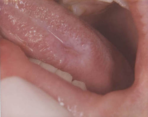
This is a 15-year-old boy who presented with a painful non-healing ulcer on the left lateral border of tongue (Figure 1). The ulcer was surrounded by a white plaque. The right buccal mucosa also had a white lesion which had a reticular pattern (Figure 2). The right buccal mucosa lesion was not ulcerated and was not painful. The left buccal mucosa had a hint of a white reticular lesion but was not as prominent as that of the right side. The ulcer was of one month’s duration while the right buccal mucosa lesion was of unknown duration. An incisional biopsy of the lateral tongue was performed.
Diagnostic Information Available
This is a 15-year-old boy who presented with a painful non-healing ulcer on the left lateral border of tongue (Figure 1). The ulcer was surrounded by a white plaque. The right buccal mucosa also had a white lesion which had a reticular pattern (Figure 2). The right buccal mucosa lesion was not ulcerated and was not painful. The left buccal mucosa had a hint of a white reticular lesion but was not as prominent as that of the right side. The ulcer was of one month’s duration while the right buccal mucosa lesion was of unknown duration. An incisional biopsy of the lateral tongue was performed.
Figure 1 This clinical photograph of the left lateral border of tongue was taken at presentation. Note the surface ulceration surrounded by a rim of white plaque.
The patient is generally in good health and has no significant past medical history. His health history is negative for diabetes. He has no history of smoking or alcohol use. He has no family history of similar lesions.
The left lateral border of tongue showed evidence of an ulcer surrounded by a rim of white plaque (Figure 1). The dorsal and right lateral border of tongue showed no evidence of similar lesions. The central part of right buccal mucosa was white and rough (Figure 2). It had white striae interlacing to produce a reticular pattern. There was no evidence of ulceration. The left buccal mucosa appeared relatively normal with a hint of white striae.
Figure 1 This clinical photograph of the left lateral border of tongue was taken at presentation. Note the surface ulceration surrounded by a rim of white plaque.
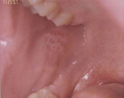
Figure 2 This clinical photograph of the right buccal mucosa was taken at presentation. Note the white striae forming a non-ulcerated reticular pattern
The biopsy material was processed for immunofluoresnce and H & E stains. The latter showed evidence of keratinized and proliferative surface epithelium with a basal cell layer exhibiting focal degeneration (Figures 3 & 4). The superficial connective tissue was infiltrated by many lymphocytes arranged in a linear fashion. The immunofluoresence stain was negative with IgG, IgA, IgM and C3 antibodies. Positive stain was identified with antibody to fibrinogen (Figure 5) present along the basement membrane in a linear fashion.
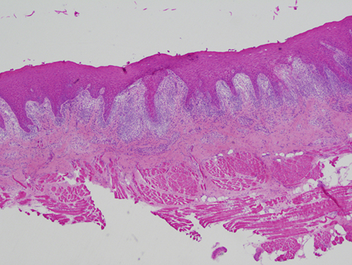
Figure 3 Screening power (40X) H & E histology shows a fragment of mucosa covered by keratinized epithelium supported by fibrous connective tissue containing a band-like infiltrate of lymphocyte.
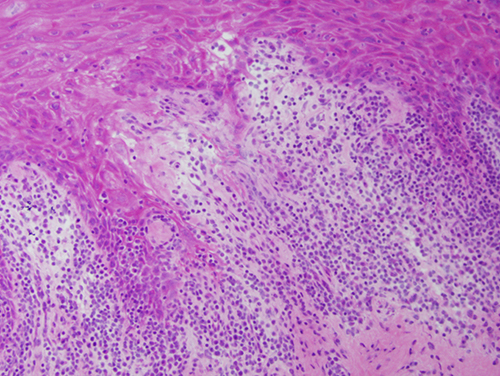
Figure 4 Higher power (200X) H & E histology of the surface epithelium exhibiting basal cell degeneration with lymphocytes arranged directly beneath the surface epithelium.
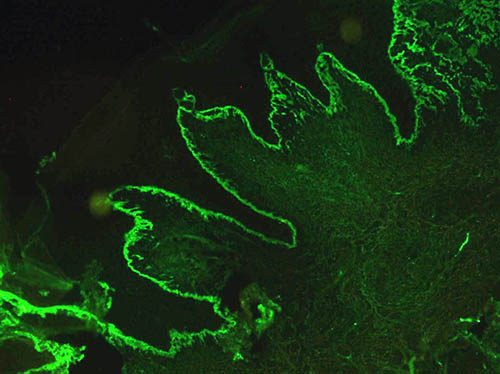
Figure 5 Low power (100X) immunofluoresence histology of the surface epithelium exhibiting a uniformly positive line of fibrinogen along the basement membrane.
After you have finished reviewing the available diagnostic information