Swelling of the Lower Anterior Jaw
Can you make the correct diagnosis?
This is a 14-year old white male who presented with a swelling of the right symphysis and parasymphysis area.
Sorry! you are incorrect
This is a benign hereditary condition that involves bilateral swelling of the maxilla and/or the mandible. Unilateral cases have been reported but are very rare. The latter are usually associated with a definitive family history of cherubism, which is autosomal dominant, and occur during childhood in patients as young as 1 year of age with an average age of 7; they slow and regress after puberty.
Patients present with a broad face due to mandibular expansion. If the maxilla is involved, the eyes may be slightly rotated upwards—hence, the name “cherubism”—due to orbital floor involvement. Diplopia is not present. Radiographically, there is multilocular radiolucency bilaterally or in all four quadrants. The histology of cherubism consists of granulation tissue stroma, giant cells and blood vessels with hyaline deposits. The radiographic changes and age of the present case are consistent with cherubism; however, the histology and lack of family history are not supportive of this condition, and the patient is not an adopted child.1
Sorry! you are incorrect
Three types of hyperparathyroidism are described: primary, which is the most common; secondary; and tertiary, which is the least common. All three types are characterized by excess parathyroid hormone production. Primary hyperparathyroidism is characterized by hypercalcemia and hypophosphatemia, secondary hyperparathyroidism is characterized by hyperphosphatemia and mild hypocalcemia, and tertiary hyperparathyroidism is characterized by hypercalcemia. Since primary hyperparathyroidism is the most common type, it will be described here in more detail than the other two types.
Primary hyperparathyroidism is usually due to an adenoma of the parathyroid gland, but is sometimes a result of hyperplasia (in about 10% of cases) and is rarely caused by adenocarcinoma.2 Secondary hyperparathyroidism is a sequelae to chronic renal disease (renal osteodystrophy). Primary hyperparathyroidism is 3 times more common in females than males and typically occurs in patients in their fifties and older. The clinical presentation is characteristically referred to as bones, stones, groans and moans—affecting multiple organs including bones, kidney stones, gastrointestinal system groans, muscles and the central nervous system moans. Bone lesions are painful multiple unilocular and multilocular radiolucencies that affect the fingers and the skull, including the jaw bones. They are called ‘brown tumors’ because of the deep chocolate brown color of the specimens resulting from hemorrhage and hemosiderin pigmentation. Stones are result of hypercalcemia affecting the kidneys and skin. Groans are related to intestinal ulcers and constipation, and moans are related to alteration in the central nervous system such as depression and sometimes seizures. This patient did not have alteration in his calcium/phosphate levels. Histologically, the biopsy material was not consistent with brown tumor of hyperparathyroidism.2
Congratulations! You are correct
Odontogenic keratocyst (OKC) was first described in 1876 and named by Phillipsen in 1956. It is an aggressive odontogenic cyst and is known for its rapid growth and its tendency to invade the adjacent tissues, including bone. It has a high recurrence rate and is associated with basal cell nevus syndrome.3,4
OKCs are derived from either the epithelial remnants of the tooth germ or the basal cell layer of the surface epithelium.3,5 The majority of patients are in the age ranges of 20-29 and 40-59, but cases in patients ranging in age from 5 to 80 years have been reported. In one study, the average age of males with OKC was 9.7 years older than that of females.4 The distribution between sexes varies from equal distribution to a male-to-female ratio of 1.6:1, except in children.3,4 OKC predominantly affects Caucasian populations and, if one may judge from the limited evidence provided by the literature, is chiefly of Northern European descent.3
Odontogenic keratocysts may occur in any part of the upper and lower jaw, with the majority (almost 70%) occurring in the mandible. They occur most commonly in the angle of the mandible and ramus.3 This is an area common to many benign odontogenic tumors such as ameloblastoma and is also a typical location for dentigerous cysts. Interestingly, the incidence of OKC seems to be increasing with time; El-Hajj has made the same observation, which he attributes to increased competence and knowledge among pathologists.6
Radiographically, OKCs present predominantly as unilocular radiolucencies with well-defined or sclerotic borders; they may also present as multilocular radiolucencies. The ratio of unilocular to multilocular OKCs varies from 3:1 to 1:1.3.7,8 Perhaps this disparity can be attributed to the fact that the multilocular appearance of OKCs is more of a unilocular radiolucency with scalloped borders lacking true compartment formation.8 Odontogenic keratocysts of the maxilla are smaller in size when compared to those occurring in the mandible; larger OKCs tend to expand bone, but mildly—obvious clinical expansion should be viewed with suspicion for a neoplasm. OKCs can also present as small and oval radiolucencies between teeth simulating a lateral periodontal cyst, in an area of an extracted tooth simulating a residual cyst, at the apex of a vital tooth mistaken for a periapical cyst, or in the anterior maxilla between the central incisors simulating an incisive canal cyst.4 OKCs grow to sizes larger than any other odontogenic cysts. They usually penetrate the bone rather than expand it and grow in an anterior to posterior direction.9 Despite this aggressive growth, they often remain asymptomatic, thus growing to large sizes and hollowing the bone.
Multiple OKCs are frequently associated with bifid-rib basal cell nevus syndrome (Gorlin syndrome). This syndrome is an autosomal dominant disorder arising from defects on chromosome 9q23.1-q31, and is often identified in juvenile kindred.10 OKC presenting as unilocular radiolucency with a sclerotic border is also known to be associated with Gorlin syndrome. Some authors suggest that as many as half of OKCs are related to Gorlin syndrome .10 When so associated, the cysts are frequently multiple and the patient is generally younger than usual, such as in the present case.9
Odontogenic keratocysts are significant clinical entities due to their tendency for recurrence and destructive behavior. They are known to have a high recurrence rate, ranging from 13% to 60%.3,4 Complete surgical removal is the treatment of choice. Surgery includes enucleation, curettage, enucleation and peripheral ostectomy, to resection depending on the radiographic presentation, location and clinical behavior.5 Surgery combined with Carnoy’s solution or liquid nitrogen treatment have been effective in reducing recurrence rate (5). At times, adjacent or associated teeth are extracted in the interest of complete removal. Some investigators advocate marsupialization and occasionally resection of the more aggressive cysts that tend to perforate buccal and lingual bone.11 Resection is a rare modality of treatment. Most cysts recur within the first three years while others may recur as late as after 16 years.3,4 Conservative surgical removal and long-term follow-up is the treatment of choice by most clinicians.
Treatment
Due to the extensive nature of the cystic lesions, they were removed in an operating room setting. Treatment included enucleation of cysts from area of teeth #s 1, 16, 17 and 9-12. The enucleated bony cavities were packed with Carnoy’s soaked gauze for approximately two minutes to cauterize any microscopic cystic epithelial tissue. The packings were removed and the bony crypts were thoroughly irrigated.
The cystic cavity in area of tooth #17 was treated in a similar manner to those in the maxilla, including Carnoy’s cauterization. The largest cyst, which was between teeth #s 21 and 31, was unroofed anterior and lateral to tooth #22 and was partially enucleated. The remaining cyst was marsupialized and was packed with approximately two 12″ long x 1″ wide Whitehead’s impregnated strips of gauze (Figure 3).
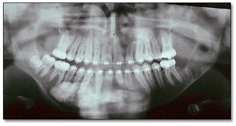
Figure 3. Panoramic post operative view of the large mandibular marsupialized cyst with radiopaque packing material.
The entire surgical site of the anterior mandible was thoroughly irrigated with sterile saline and partially closed with silk suture in an interrupted fashion. A window of approximately 1″ was left open for irrigation and packing removal (Figure 4). The packing was changed every 2-3 weeks for a total of 8-9 months.
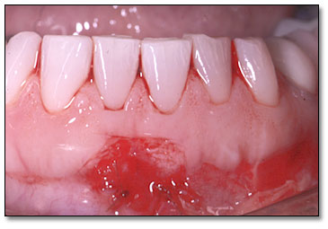
Figure 4. Anterior mandible demonstrating partially closed surgical site with a window left open for irrigation and packing removal.
The sixth OKC in the area of impacted tooth #32 was also enucleated and Carnoy’s cauterized in a manner similar to all the other cysts. A total of three teeth were extracted (teeth #s 1, 17 and 32), and five teeth were endodontically treated (teeth #s 23-27) (Figure 5). In addition, the patient manifested features of mild hypertelorism, frontal bulging, sunken eyes, and heavy and fused eyebrows consistent with bifid rib basal cell nevus syndrome.
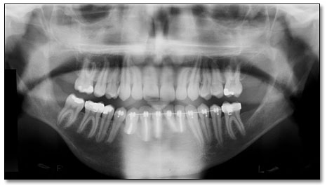
Figure 5. Approximately 18 month post operative panoramic radiograph, demonstrating complete healing of the surgical sites of all six cysts.
References
- Ozkan Y, Varol A et al. Clinical and radiological evaluation of cherubism: a sporadic case report and review of the literature. Int J Pediatr Otorhinolaryngol. 2003; 67:1005-1012.
- Yamazaki H, Ota Y et al. Brown tumor of the maxilla and mandible: progressive mandibular brown tumor after removal of parathyroid adenoma. J Oral Maxillofac Surg. 2003; 61:719-722.
- Shear M. Odontogenic keratocysts: natural history and immunohistochemistry. Oral Maxillofacial Surg Clin N Am. 2003; 15: 347-362.
- Oda D, Rivera V et al. Odontogenic keratocyst: the northwestern USA experience. J Contemp Dent Pract. 2000 Feb 15; 1(2): 60-74.
- Stoelinga, PJW. Excision of the overlying, attached mucosa, in conjunction with cyst enucleation and treatment of the bony defect with carnoy solution. Oral Maxillofacial Surg Clin N Am. 2003; 15: 407-414.
- El-Hajj G, Anneroth G. Odontogenic keratocysts – a retrospective clinical and histological study. Int J Oral Maxillofac Surg. 1996; 25: 124-29.
- Haring JI, Van Dis ML. Odontogenic keratocysts; a clinical, radiographic and histopathologic study. Oral Surg Oral Med Oral Pathol. 1988; 66: 145-153.
- Chen C-H, Lin C-C. Clinical and histopathological study of the odontogenic keratocyst – a follow-up study of 16 cases. Kaohsiung J Med Sci. 1986; 2: 601-607.
- Zachariades N, Papanicolaou S, Triantafyllou D. Odontogenic keratocysts: Review of the literature and report of sixteen cases. J Oral Maxillofac Surg. 1985; 43: 177-182.
- Fardon PA, Norris D.J et al. Analysis of 133 meioses places the genes for nevoid basal cell carcinoma (Gorlin) syndrome and Fanconi anemia group C in a 2.6-cM interval and contributes to the fine map of 9q22.3, Genomics. 1994; 23: 486-489.
- Irvine GH, Bowerman JE. Mandibular keratocyst: Surgical management. Br J Oral Maxillofac Surg. 1985; 23: 203-209.