Return to Case of the Month Archives
Fast-growing soft tissue lesion posterior mandible
Dolphine Oda, BDS, MSc
doda@u.washington.edu
Contributed by
Drs. Patricia Kelly and Mark Egbert
University of Washington and Seattle Children’s Hospital
Case Summary and Diagnostic Information
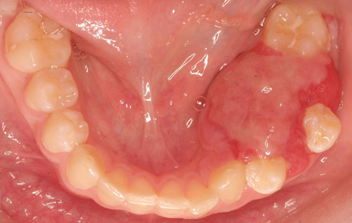
This is a 13-year old white male who presented with a rapidly growing swelling of the left posterior mandible (Figures 1), area of teeth #s 19-21 of one month’s duration with significant displacement of teeth and completely covering tooth # 20.
Diagnostic Information Available
This is a 13-year old white male who presented with a rapidly growing swelling of the left posterior mandible (Figures 1), area of teeth #s 19-21 of one month’s duration with significant displacement of teeth and completely covering tooth # 20. There was no history of trauma, purulence or bleeding. The lesion was asymptomatic including no sensory disturbances. The panoramic view revealed a large and well circumscribed radiolucency centered over tooth #20 and displacing teeth #s19-21 (Figure 2).
Figure 1 Occlusal view. Photograph taken at first clinical presentation to Children’s Hospital. Note the pink/red, sessile swelling expanding the mandible both buccal and lingual displacing teeth #s 19 and 21. Lingual expansion is more prominent. Tooth # 20 is completely covered by the neoplasm
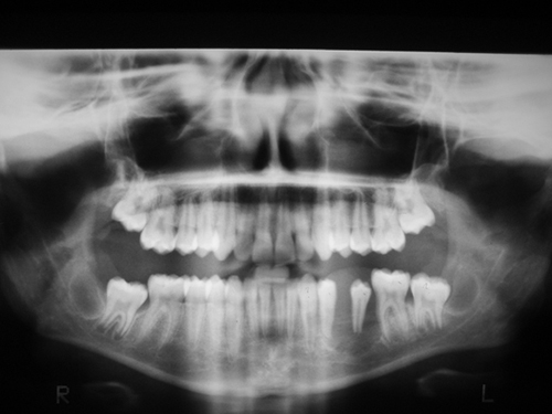
Figure 2 Panoramic radiograph demonstrating bone resorption, left mandible in the area of teeth # 19-21. This radiograph reveals a well-demarcated destructive soft tissue lesion. The bone in the area is almost all resorbed giving the impression of teeth floating in space
The past medical history is unremarkable and the family history is non contributory.
Clinical examination revealed a firm, pink, fleshy mass in the left posterior mandible with intact surface and displaced teeth numbers 19, 20, and 21 (Figures 1). Multiple radiographic images—panoramic (Figure 2), and computed tomography (CT) scan (Figure 3) were taken and all revealed a destructive soft tissue lesion with significant buccal and lingual expansion. An incisional biopsy was performed. Once the diagnosis was obtained, the patient was planned for an excisional biopsy of the entire lesion.
Figure 1 Occlusal view. Photograph taken at first clinical presentation to Children’s Hospital. Note the pink/red, sessile swelling expanding the mandible both buccal and lingual displacing teeth #s 19 and 21. Lingual expansion is more prominent. Tooth # 20 is completely covered by the neoplasm.
Figure 2 Panoramic radiograph demonstrating bone resorption, left mandible in the area of teeth # 19-21. This radiograph reveals a well-demarcated destructive soft tissue lesion. The bone in the area is almost all resorbed giving the impression of teeth floating in space.
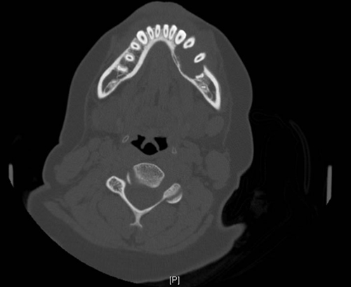
Figure 3 Computed tomography (CT) scan image reveals a destructive soft tissue lesion with significant buccal and lingual expansion.
Treatment
Under general anesthesia, the lesion was completely excised along with teeth # 19-21 in one en bloc mass. Iodoform gauze packing was sutured into the defect for 10 days (Figure 4). His postoperative regimen included wound care and observation only. Post-operative healing was unremarkable. After a period of one month, the site appears well healed. The patient will continue to be followed, and planned in the future (after completion of growth) for bone grafting and eventually for implant prothesis.
Incisional and excisional biopsy
Histological examination revealed a highly cellular fibrous lesion with focal myxoid background. The fibroblasts are eosinophilic with plump and vesicular nuclei and are arranged in elongated fascicles with focal areas of storiform pattern (Figures 5 & 6). Mitotic figures are present but are scant. There is no evidence of pleomorphism. The immunohistochemistry markers are positive with smooth muscle marker HHF35, but negative with desmin, myogenin, and S-100 proteins.
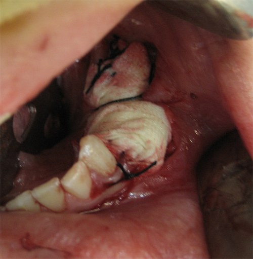
Figure 4 Surgical site packed with iodoform gauze and sutured for ten days.
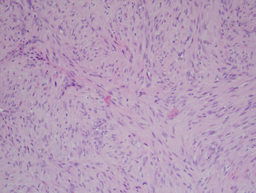
Figure 5 Low power (x100) histology shows short bundles of spindle-shaped fibroblasts criss-crossing each other, some in a storiform pattern. There was no evidence of atypia and mitotic figures were occasional to absent.
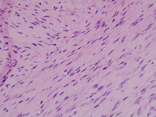
Figure 6 High power (x200) histology shows short bundles of spindle-shaped fibroblasts criss-crossing each other. The neoplastic cells show no evidence of atypia.
After you have finished reviewing the available diagnostic information