Slowly Enlarging Lesion on the Right Lateral Tongue
Can you make the correct diagnosis?
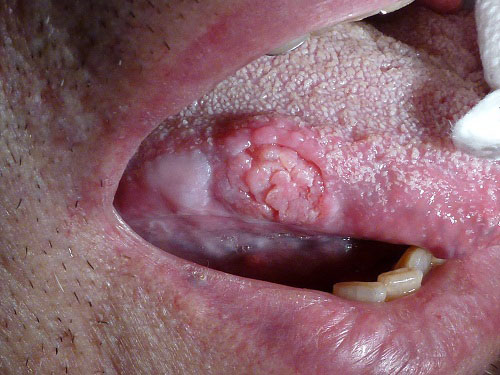
This is an 82-year old white male with a two-year history of a slowly enlarging lesion on the right lateral border of tongue.
Sorry, you are incorrect!
Although the center of this lesion is not frankly ulcerated, the rolled border suggests that may have been ulcerated. The site and the age of the patient are consistent with traumatic ulcerative granuloma with stromal eosinophilia (TUGSE). The duration of two years is not typical, neither is the gender. The histology was not supportive of TUGSE.
Traumatic ulcerative granuloma with stromal eosinophilia (TUGSE) ulcers are also known by other names, including eosinophilic ulcer, ulcerative eosinophilic granuloma of the tongue (which is not related to eosinophilic granuloma of the bone as in Langerhans Cell Histiocytosis), and traumatic granuloma. Traumatic ulcerative granuloma with stromal eosinophilia commonly occurs on the dorsal and lateral tongue, usually in patients with a history of trauma. The latter is not always clearly demonstrated. Although the tongue is the most common location, TUGSE has also been described in other areas such as the lips, buccal mucosa, gingiva and vestibule. TUGSE affects patients of all ages but is more common in adult males. It is described in children as young as infants with Riga-Fede, a disease that occurs during the first year of life as a result of chronic trauma in the sublingual area and ventral tongue occurring during breastfeeding due to the sharp-edged central lower incisors. The clinical presentation of TUGSE ranges from a small ulcer with rolled borders to an exophytic large lesion simulating a pyogenic granuloma. It can also present as a very large, indurated and deep ulcer simulating a squamous cell carcinoma or any other malignancy of the mouth. The duration of the non-healing ulcer can range from two weeks to many months. TUGSE tends to occur more in males than females. Histologically, these ulcers are deep lesions involving the underlying muscle, which may explain the process of slow healing and the tissue eosinophilia. Healing may take up to eight weeks, sometimes more.
Congratulations, you are correct!
Although the bluntly papillary surface is not a typical clinical presentation of oral squamous cell carcinoma, the rolled border, the duration, the risk factor of tobacco use (although in the distant past) are all consistent with oral SCC.
Oral squamous cell carcinoma is a highly aggressive neoplasm that currently ranks as the fifth most common malignant neoplasm worldwide and accounts for an estimated 90% of all oral malignancies. It is predominantly diagnosed in males over the age of 40 years, with an observed male-to-female ratio of 2:1 generally and 1.4:1 in the United States. The incidence of oral SCC in patients under the age of 40 is relatively low, around 5%. Prolonged histories of tobacco and alcohol consumption are the main risk factors in both populations. Other risk factors including human papilloma virus, especially type 16, have been reported to be significant, especially in the young male population with tonsillar and laryngeal squamous cell carcinomas. Other factors include poor oral hygiene, syphilis, chronic candidiasis, iron and dietary deficiencies, herpes simplex and various other immunologic factors, and lichen planus—especially persistent erosive lichen planus. The most common sites of occurrence for oral squamous cell carcinoma are the tongue, the ventral and lateral surfaces, and the floor of mouth. Excluding the outer lip, the most common sites (in decreasing order) are the ventral and lateral surfaces of the tongue (25-50%), floor of mouth (15%), gingiva (12%) and palate (9%). The buccal mucosa and retromolar pad areas (3%) have a relatively low incidence of occurrence unless the patient is a chronic smokeless tobacco user. Oral SCC varies in presentation from deceptively benign looking to obviously malignant. It may present as a non-healing ulcer, or as red, white or mixed red-and-white lesions. Characteristic signs of oral SCC are non-healing ulcer, ulcer with rolled borders, fungation, fixation and induration. Rarely, oral SCC may present as unexplained asymptomatic lateral neck lymphadenopathy. Determination of the prognosis of oral SCC is based on its clinical stage and histological classification. Although oral SCC is a diagnosis made by histology, surgeons tend to depend exclusively on the TNM classification system for clinical staging and treatment decisions. Prognosis is dependent on the TNM staging system; the most important prognostic sign is the presence or absence of metastases at the time of diagnosis. The prognosis thus improves when the lesion is detected early. Oral SCC patients die mainly of infection due to lowered resistance or of hemorrhage if the tumor erodes through one of the main blood vessels.
References
- Eleni G, Panagiotis S, Andreas K, Georgia A. Traumatic ulcerative granuloma with stromal eosinophilia: a lesion with alarming histopathologic presentation and benign clinical course. Am J Dermatopathol. 2011 Apr;33(2):192-4.
- Boffano P, Gallesio C, Campisi P, Roccia F. Traumatic ulcerative granuloma with stromal eosinophilia of the retromolar region. Craniofac Surg. 2009 Nov;20(6):2150-2.
- Hirshberg A, Amariglio N, Akrish S, Yahalom R, Rosenbaum H, Okon E, Kaplan I. Traumatic ulcerative granuloma with stromal eosinophilia: a reactive lesion of the oral mucosa. Am J Clin Pathol. 2006 Oct;126(4):522-9.
- Bundgaard T, SM Bentzen, J Wildt, FB Sorensen, H Sogaard and JE Nielsen, Histopathologic, stereologic, epidemiologic, and clinical parameters in the prognostic evaluation of squamous cell carcinoma of the oral cavity, Head & Neck 18 (1996), pp. 142–152. Full Text via CrossRef | View Record in Scopus | Cited By in Scopus (52)
- El-Mofty SK and DW Lu, Prevalence of human papillomavirus type 16 DNA in squamous cell carcinoma of the palatine tonsil, and not the oral cavity, in young patients: a distinct clinicopathologic and molecular disease entity, Am J Surg Pathol 27 (2003) (11), pp. 1463–1470. Full Text via CrossRef | View Record in Scopus | Cited By in Scopus (60)
- Scully C, Oral cancer; the evidence for sexual transmission, Br Dent J 199 (2005), pp. 203–207. Full Text via CrossRef | View Record in Scopus | Cited By in Scopus (23)
- Manuel S, SK Raghavan, M Pandey and P Sebastian, Survival in patients under 45 years with squamous cell carcinoma of the oral tongue, Int J Oral Maxillofac Surg 32 (2003)
- Mathew Iype, M Pandey, A Mathew, G Thomas, P Sebastian and M Krishnan Nair, Squamous cell carcinoma of the tongue among young Indian adults, Neoplasia 3 (2001), pp. 273–277.
- Gharebaghi N, Monsouri SA, Darazam IA, Mansouri D, Sajadi MM, Mansouri N. A 40-year-old man with tongue lesions. Lingual and pulmonary tuberculosis (TB). Clin Infect Dis. 2011 May;52(10):1231, 1276-7.
- Bernstein JM, Bacheller CD. It’s on the tip of my tongue. Skinmed. 2006 May-Jun;5(3):142-5.
- Akin L, Herford AS, Cicciù M. Oral presentation of disseminated histoplasmosis: a case report and literature review. J Oral Maxillofac Surg. 2011 Feb;69(2):535-41. Epub 2010 Dec 9.
Sorry, you are incorrect!
The site of the lesion, the rolled border, the bluntly papillary surface are all consistent with the presentation of a chronic granulomatous type of condition be it infectious or of other etiology. The duration of two years is not supportive of an infectious condition but can be a foreign body granuloma type of granulomatous process. The histology was not supportive of infectious or other granulomatous processes.
Some infections of the oral cavity, especially the deep granulomatous type of infectious diseases, may present as diffuse, deep ulcers with rolled borders on the lateral ventral tongue comparable to the clinical presentation of malignant neoplasms such as squamous cell carcinoma.
The oral cavity is a site where a number of acute and chronic infectious diseases occur as a local primary disease or as a manifestation of a systemic disease. They include diseases of viral, fungal and bacterial origin. They usually present in multiples or in a diffuse manner ranging from ulcers to small and nodular lesions to verrucoid lesions. Occasionally, granulomatous-type infectious diseases present as diffuse and deeply ulcerative lesions with rolled borders, simulating oral SCC. These cases include ulceration induced by mycobacterium tuberculosis, deep fungal infections such as blastomycosis and Histoplasmosis, and chancre of primary syphilis. Oral manifestations of tuberculosis and Histoplasmosis are unusual without lung involvement and a disseminated disease; they occur more often in immune-compromised patients. The gingiva and the mandibular vestibule are more common locations, but the tongue can occasionally be affected. Chancre can occur in any location, including the tongue, lips and hard palate.