Single smooth surfaced and well-demarcated nodule, ventral tongue
Can you make the correct diagnosis?
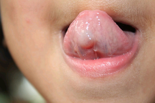
This is a healthy four-year-old girl whose mother was the first to notice a single, smooth-surfaced, well-demarcated and grayish-pink nodule on the right side of the ventral surface of tongue about 4 X 4 mm in size.
Sorry! you are incorrect
Given the location and the age of the patient, Lymphangioma should be on the differential diagnosis. However, the histology is not supportive of Lymphangioma.
Lymphangioma is a benign congenital vascular developmental anomaly of the lymphatic system that commonly occurs in the head and neck area, including the oral cavity. It is best classified as being either superficial or deep, with the latter divided into cavernous and cystic (cystic hygroma). The superficial type is also known as lymphangioma simplex (1-3). Oral lymphangiomas tend to be of the cavernous type. The head and neck area is the most common location for lymphangiomas, followed by the extremities and buttocks. Up to 50% of lymphangiomas occur at birth and almost 90% develop within the first two years of life (1-3). In the oral cavity, lymphangiomas most commonly affect the tongue; if superficial, it has a "cobblestone" appearance with clusters of fluid filled vesicles, some with dark red color due to bleeding in the area. If it is deep, the lesion will be more diffuse and soft in consistency (1-3). It may also occur on the lips, resembling an angioedema. It is usually asymptomatic, but it can be painful if pressed and it may drain clear fluid if traumatized.
Histologically, lymphangiomas consist of dilated lymphatic vessels present directly beneath the surface epithelium, some extending into the underlying deep connective tissue. Surgery, scalpel or CO2 laser are the preferred treatments for this lesion. Sclerosing agents such as OK-432 and steroids have been used with some success (2).; Local recurrence is common, especially with the deep and hard-to-reach lesions. The superficial lesions have better success and fewer incidences of recurrence.
Congratulations! You are correct
Ventral tongue mucoceles are very rare, and for that reason they were not high on the clinical differential diagnosis. Mucoceles and ranulas are clinical terms describing exophytic, fluid filled, fluctuant nodules, typically of minor salivary gland origin and present mostly on the lower lip and the floor of the mouth (4-6). Over 90% of these lesions are cyst-like structures, or pseudocysts, and are mucous extravasation phenomena referred to as mucoceles. Some of these lesions are true cystic structures lined by epithelium and filled with mucus and are called mucus retention cysts or salivary duct cysts; these constitute a small percentage of all mucoceles (4-6). Ranulas, mucoceles of the floor of the mouth, constitute the other 5% and are divided into those above the mylohyoid muscle (majority) and below the mylohyoid muscle (also known as plunging ranulas or cervical ranulas) (7-8). Ranulas are of minor or major salivary gland origin and are mostly extravasation in type. The etiology of the extravasation mucoceles is usually sharp trauma cutting through the salivary gland duct and releasing the mucous in the extracellular tissue (4-6). Histologically, the extravasation-type mucocele consists of a cyst-like structure lined by granulation tissue and filled with mucoid material, foamy macrophages, and at times small clusters of neutrophils. The mucous retention cysts develop as a result of a duct blockage which can be caused by trauma, fibrosis, sialolith, or pressure from an overlying tumor (5-6). The extravasation mucoceles most commonly occur on the lower lip and very rarely on upper lip. They may occur anywhere else in the oral cavity, including the buccal mucosa and floor of mouth (Ranula). The latter can be of minor salivary gland or submandibular or sublingual gland duct origin (7-8). It is more commonly seen in children and adolescents. It presents as a swelling with a bluish color if superficial, while deep mucoceles (like this case) tend to take the color of the surrounding mucosa. Mucoceles tend to fluctuate in size. They are usually associated with a history of sharp lip or cheek biting, but can also be secondary to surgery in the area. This is especially true with the anterior tongue mucoceles. Surgical excision with the associated minor salivary gland is the preferred treatment for the deep mucoceles; superficial mucoceles can self-heal within 2-3 weeks. Superficial mucoceles can also mimic vesiculobullous-type diseases because they look like vesicles (5), especially when presenting in multiples (rare, but described). They can recur if the source of trauma is not eliminated or if they are secondary to surgery. Simple (non-plunging) ranula is best treated by marsupialization into the floor of mouth (8). Plunging ranula requires complete excision via an extra-oral approach. The technical difficulties associated with the complete removal of this thin-walled lesion result in a relatively high recurrence rate.
Treatment
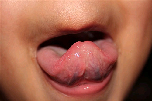
Under general anesthesia, the lesion was completely excised, upon which point it was described as having a fluid filled yellow to honey-like color with fluid emerging from the central cystic structure. The surgical site was closed with Vicryl suture. The area healed well within two weeks; however, a small and superficial mucoceles appeared in the third week, lasted for two weeks and healed without surgery. The clinical presentation five months after surgery (Fig 2) shows a completely healed area with very little scarring.
References
- Jian XC. Surgical management of lymphangiomatous or lymphangiohemangiomatous macroglossia. J Oral Maxillofac Surg. 2005 Jan;63(1):15-9.
- Wheeler JS, Morreau P, Mahadevan M, Pease P. OK-432 and lymphatic malformations in children: the Starship Children’s Hospital experience. ANZ J Surg. 2004 Oct;74(10):855-8.
- Al-Salem AH. Lymphangiomas in infancy and childhood. Saudi Med J. 2004 Apr;25(4):466-9.
- Kapoor N, Bhalla J, Bharadwaj VK, Kotgirwar BK. Cartilagenous choristoma of palatine tonsil–a case report. Indian J Pathol Microbiol. 2003 Oct;46(4):654-5.
- Toida M, Sugiyama T, Kato Y. Cartilaginous choristoma of the tongue.
J Oral Maxillofac Surg. 2003 Mar;61(3):393-6. Review. - Mintz S, Anavi Y, Barak S, Nalley K. Osseous choristomas of the buccal mucosa. J Mich Dent Assoc. 1995 Mar;77(3):30-2, 55. Review
- Kopp WK, St-Hilaire H. Mucosal preservation in the treatment of mucocele with CO2 laser. J Oral Maxillofac Surg. 2004 Dec;62(12):1559-61.
- Silva A Jr, Nikitakis NG, Balciunas BA, Meiller TF. Superficial mucocele of the labial mucosa: a case report and review of the literature. Gen Dent. 2004 Sep-Oct;52(5):424-7.
- Auclair PL, Ellis GL. Mucoepidermoid carcinoma. In Ellis GL, Auclair PL, Gnepp DR, editors. Surgical pathology of the salivary glands. Philadelphia: W.B. Saunders, 1991. p. 269-298.
- Hicks J, Flaitz C. Mucoepidermoid carcinoma of salivary glands in children and adolescents: assessment of proliferation markers. Oral Oncol. 2000 Sep;36(5):454-60
- Bentz BG, Hughes CA, Ludemann JP, Maddalozzo J. Masses of the salivary gland region in children. Arch Otolaryngol Head Neck Surg. 2000 Dec;126(12):1435-9.
- Epstein JB, Hollender L, Pruzan SR. Mucoepidermoid carcinoma in a young adult: recognition, diagnosis, and treatment and responsibility. Gen Dent. 2004 Sep-Oct;52(5):434-9.
- Fletcher CDM. Soft tissue tumors. In: Fletcher CDM, editor. Diagnostic histopathology of tumors, 2nd edition (Volume 2). London: Churchill Livingstone, 2000. p. 1473-1540.
- Fernandez JM, Medlich MA, Lopez LH, Loredo CF, Luque LC. Fetal intermediate rhabdomyoma of the lip: case report. J Clin Pediatr Dent. 2005 Winter;29(2):179-80
- O’Callaghan MG, House M, Ebay S, Bhadelia R. Rhabdomyoma of the head and neck demonstrated by prenatal magnetic resonance imaging. J Comput Assist Tomogr. 2005 Jan-Feb;29(1):130-2.
- Xue JL, Fan MW, Wang SZ, Chen XM, Li Y. A clinicopathological study of 14 cases of oral granular cell tumor
- Brannon RB, Anand PM. Oral granular cell tumors: an analysis of 10 new pediatric and adolescent cases and a review of the literature. J Clin Pediatr Dent. 2004;29(1):69-74.
Sorry! you are incorrect
Given the grayish color and fluctuant nature of the early presentation, mucous producing neoplasms should be included on the differential diagnosis. The histology, however, is not supportive of this diagnosis.
Mucoepidermoid carcinoma is a malignant neoplasm of salivary gland origin that can present as a smooth-surfaced swelling or a non-healing ulcer on the palate. It occurs in a wide age range (9-11). Three histologic types are reported: low, intermediate and high; the low-grade type is more common in the oral cavity (9). Mucoepidermoid carcinoma accounts for 10% of all salivary gland neoplasms (9-12). While the majority occurs in the parotid gland, some also occur in minor salivary glands, especially the palate, tongue, buccal mucosa, lips, and retromolar pad areas (9-12). It can occur at any age with a predilection for young people (9). The Armed Forces Institute of Pathology (AFIP) studies demonstrate 44% of cases occurring in patients under 20 years of age, most commonly on the palate (9-10). Their youngest patient was nine months old. The low-grade lesions are slow-growing and painless, and not encapsulated; they sometimes resemble a mucocele, especially those at the retromolar pad area (10-11). Retromolar pad area mucoceles are rare, and for that reason it is best to biopsy early to exclude the possibility of a mucoepidermoid carcinoma masquerading as a mucocele. High-grade lesions tend to be more common in the parotid gland; they present as rapidly growing, painful lesions with facial nerve paralysis and sometimes with regional lymph node metastasis. Histologically, mucoepidermoid carcinoma consists of a variety of cell types and architectural patterns which constitute the three histologic gradings. Although low-grade mucoepidermoid carcinoma is characterized by an abundance of mucous-producing cells and duct-like structures with cystic dilation, the mere presence of certain types of cells and architecture should not be used to determine the histologic grade. The full discussion of the histology of this lesion is beyond the scope of this chapter. Complete surgical removal with clean margins is the preferred treatment for the low-grade type. Radiotherapy has also been successfully used, especially when the tumor involves the surgical margins (9-12).
Sorry! you are incorrect
The location, age and firm consistency is supportive of this lesion. However, the histology is not.
This is a benign and very rare neoplasm of skeletal muscle origin. It can be misdiagnosed as embryonal rhabdomyosarcoma (13, 15). The head and neck area is the most common location for fetal rhabdomyomas, especially behind the ears and the tongue (13). It occurs predominantly in children and is slow-growing and rarely recurs (13-15). Histologically, it can be myxoid or cellular and it can have mature rhabdomyoblasts without evidence of atypia or mitosis (14). Treatment includes conservative surgical excision.
Sorry! you are incorrect
The location is somewhat supportive. However, the age and the histology are not. Granular cell tumors tend to occur on the dorsal lateral tongue.
This is a benign neoplasm of nerve origin supported by Immunohistochemistry markers (16). The tongue is the most common site of occurrence of this tumor; almost one third of cases occur in the tongue, more dorsal-lateral than ventral. Although skin is another common site, this lesion can occur in a variety of sites and tends to be benign in most cases. A malignant form is described, but it is rare.
In the mouth, the buccal mucosa is second to the tongue in site predilection, and the lesion is more common in females than males (2:1 ratio). Although it tends to occur in adults over 30 years of age, it has also been described in children of an average age of 14.5 in a 3-19 range (17). In children it is 3:1 females and has a 50% occurrence in the tongue (17). It is usually asymptomatic and can be of a long-term duration, ranging from months to years. The dorsal-lateral tongue is a common location; in about 80% of cases it is superficial and submucosal. The color ranges from pink or white with a keratotic surface to yellow-orange. A newborn counterpart is described and is known as congenital epulis of the newborn; it is believed to be a separate entity with different cell origin (16-17).
Histologically, granular cell tumor is composed of strands and fascicles of large cells with distinct cell borders containing abundant granular cytoplasm. The nuclei are small and round to oval and are eccentrically located. These cells are at times intimately related to surrounding skeletal muscle fibers and at times to nerve. The lesion can be well demarcated and confined or infiltrative. The overlying epithelium can be normal in thickness or proliferative with psuedoepitheliomatous hyperplasia (PEH) (16-17). The latter, when extensive in rare cases, can be mistaken for well differentiated squamous cell carcinoma, especially if the biopsy is superficial.
The tumor cells are positive with S-100 protein and neuron specific enolase (NSE) indicating a neural crest origin. Treatment includes conservative surgical excision. Recurrence is extremely rare.