August 2009: Swelling right posterior hard palate
Can you make the correct diagnosis?
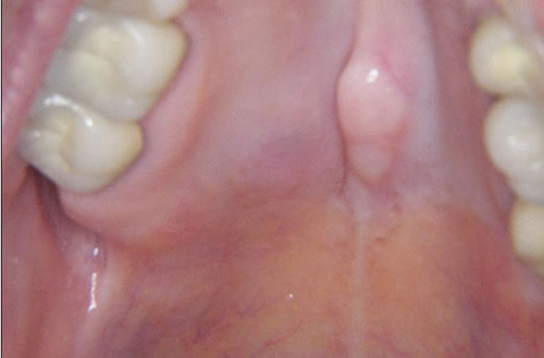
This is a 58-year-old white female with a small and slightly raised swelling on the posterior hard palate of two months’ duration (Figure 1). The swelling is asymptomatic and is soft in consistency with an intact mucosa; it is 1 x 1 cm at its greatest dimensions. It is pink to focally light blue. This patient has palatal torus that has been present for many years; this lesion is to the right of the palatal torus.
Sorry! you are incorrect
This patient has torus platinus and it has been present for many years. The lesion is right of the torus and is slightly raised and focally light blue. The soft consistency is not supportive of torus paltinus, neither is the histology.
Torus palatinus is a hyperplastic cortical bone swelling located in the midline of the hard palate and is hard on palpation. Patients usually give a history of its long term (many years’) presence. In one study it affected up to 38.5% of Asian females and 30% of white females (1-3). The etiology of this lesion is unknown, but heredity plays a role; some cases follow an autosomal dominant pattern of transmission. Torus palatinus is a bony outgrowth at the midline of the hard palate covered by normal, sometimes thin, mucosa; at times, the mucosa undergoes ulceration (1-3). It can be smooth surfaced or multilobular, in which case it usually grows in four coalescing bony lobules. It is a slow-growing and painless lesion except if traumatized. The lesion occurs by age 30 and affects females twice as often as males. Larger lesions can interfere with speech, hygiene and dental prostheses. Radiographically, it is always radiopaque. Histologically, it is made up of dense viable bone with Haversian systems containing viable osteocytes (1-3). Treatment is only indicated if it is symptomatic, interfering with function or creating difficulty with prostheses. Treatment is also indicated in patients who are about to receive Fosamax or any other type of bisphosphonates especially if they are to be taken on long-term basis and if the torus palatines is large in size (3). These lesions are most easily removed by reflection of a mucoperiosteal flap and burring away with a hand piece.
Sorry! you are incorrect
The age, location and gender are consistent with pleomorphic adenoma (PA). Given the fact that PA is the most common salivary gland neoplasm makes it important to be considered on the differential diagnosis of a palatal swelling. The color and duration however, are not supportive of PA, neither is the histology.
Pleomorphic adenoma is the most common benign salivary gland neoplasm of both the major and minor salivary glands. It originates from the myoepithelial cells and the reserve cells of the intercalated ducts. It accounts for 80% of all benign salivary gland neoplasms. It occurs in both major and minor salivary glands and accounts for up to 77% of parotid, 68% of submandibular, and 43% of minor salivary gland tumors (4-6). It is most common in females 30-50 years of age, but it is also described in children (4). One study reports 1% of cases affecting children under 10 years of age and 5.9% between the ages of 10-20 (4, 6). It presents as a small, painless, slowly enlarging nodule. If left untreated, it can enlarge significantly, sometimes increasing by several pounds in weight (1-4). It occurs in the oral cavity, especially the palate and lips (4-6). On the palate, it is usually located in the posterior hard palate or anterior soft palate but can also be in the posterior soft palate; PA usually occurs in the posterior and lateral palate, as opposed to torus palatines, which usually occurs in the middle hard palate and in the anterior. The posterior hard palate mixed tumor is fixed due to the bone-bound anatomy of the region; the tumor is otherwise movable. Histologically, mixed tumor has a wide variety of cellular and pattern manifestations; the main cellular components are epithelial duct-like structures and mesenchymal-like tissue such as myxochondroid matrix. These lesions are generally encapsulated, ranging from predominantly myxoid (36%) to extremely cellular (12%) (4-6). Complete surgical removal with clean margins is the preferred treatment (4-6). Palatal lesions respond well to excision in one piece with the periosteum and overlying mucosa. PA has a good prognosis, but it has a tendency for recurrence (up to 44%) if not treated thoroughly (1-4). Risk of recurrence is less if it occurs in the minor salivary glands (up to 20%). The risk of malignant transformation is about 5% (4, 6).
Congratulations! You are correct
Mucoepidermoid carcinoma is a malignant neoplasm of salivary gland origin that can present as a smooth-surfaced swelling or a non-healing ulcer on the palate. It occurs in a wide age range (7-9). Three histologic types are reported: low, intermediate and high; the low-grade type is more common in the oral cavity (7). Mucoepidermoid carcinoma accounts for 10% of all salivary gland neoplasms (7-9). While the majority occur in the parotid gland, some also occur in minor salivary glands, especially the palate, tongue, buccal mucosa, lips, and retromolar pad areas (7-9). It can occur at any age with a predilection for occurrence in young people (7). Studies by the Armed Forces Institute of Pathology (AFIP) find 44% of cases occurring in patients under 20 years of age, most commonly on the palate (7). Their youngest patient was nine months old. The low-grade lesions are slow-growing and painless, and not encapsulated; they sometimes resemble a mucocele, especially those at the retromolar pad area (7-9). Retromolar pad area mucoceles are rare, and for that reason it is best to biopsy early to exclude the possibility of a mucoepidermoid carcinoma masquerading as a mucocele. High-grade lesions tend to be more common in the parotid gland; they present as rapidly growing, painful lesions with facial nerve paralysis and sometimes with regional lymph node metastasis. Histologically, mucoepidermoid carcinoma consists of a variety of cell types and architectural patterns which constitute the three histologic gradings. Although low-grade mucoepidermoid carcinoma is characterized by an abundance of mucous-producing cells and duct-like structures with cystic dilation, the mere presence of certain types of cells and architecture should not be used to determine the histologic grade. Complete surgical removal with clean margins is the preferred treatment for the low-grade type. Radiotherapy has also been successfully used, especially when the tumor involves the surgical margins (7-9).
References
- Sisman Y, Ertas ET, Gokce C, Akgunlu F. Prevalence of torus palatinus in cappadocia region population of Turkey. Eur J Dent. 2008 Oct;2(4):269-75.
- Sisman Y, Gokce C, Tarim Ertas E, Sipahioglu M, Akgunlu F. Investigation of elongated styloid process prevalence in patients with torus palatinus. Clin Oral Investig. 2008 Oct 30.
- Goldman ML, Denduluri N, Berman AW, Sausville R, Guadagnini JP, Kleiner DE, Brahim JS, Swain SM. A novel case of bisphosphonate-related osteonecrosis of the torus palatinus in a patient with metastatic breast cancer. Oncology. 2006;71(3-4):306-8. Epub 2007 Jul 26.
- Waldrom CA. Mixed tumor (pleomorphic adenoma) and myoepithelioma. In Ellis GL, Auclair PL, Gnepp DR, editors. Surgical pathology of the salivary glands. Philadelphia: W.B. Saunders, 1991. p. 165-186.
- Foote FW Jr., Frazell EL. Tumors of the major salivary glands. Atlas of tumor pathology, Section IV, Fascicle 11, 1st Series. Washington DC: Armed Forces Institute of Pathology, 1954.
- Bablani D, Bansal S, Shetty SJ, Desai R, Kulkarni SR, Prasad P, Karjodkar FR. Pleomorphic adenoma of the cheek: a case report and review. J Oral Maxillofac Surg. 2009 Jul;67(7):1539-42.
- Auclair PL, Ellis GL. Mucoepidermoid carcinoma. In Ellis GL, Auclair PL, Gnepp DR, editors. Surgical pathology of the salivary glands. Philadelphia: W.B. Saunders, 1991. p. 269-298.
- Hicks J, Flaitz C. Mucoepidermoid carcinoma of salivary glands in children and adolescents: assessment of proliferation markers. Oral Oncol. 2000;36(5):454-60
- Brandwein MS, Ivanov K, Wallace DI, Hille JJ, Wang B, Fahmy A, Bodian C, Urken ML, Gnepp DR, Huvos A, Lumerman H, Mills SE. Mucoepidermoid carcinoma: a clinicopathologic study of 80 patients with special reference to histological grading. Am J Surg Pathol 2001; 25:835-45.
- Kämmerer PW, Kreft A, Toyoshima T, Al-Nawas B, Klein MO. Misleading initial histological diagnosis of a polymorphous low-grade adenocarcinoma in situ ex pleomorphic adenoma-a case report. Oral Maxillofac Surg. 2009 Jun;13(2):99-103.
- Wei YC, Huang CC, Chien CY, Hwang JC, Chen WJ. Polymorphous low-grade adenocarcinoma of the nasopharynx: a case report and brief review. J Clin Pathol. 2008 Oct;61(10):1124-6.
- Fletcher CDM. Diagnostic histopathology of tumors. Volume 1, 3rd edition; 2007. Pages 288-291.
Sorry! you are incorrect
This is a common low-grade malignant neoplasm of minor salivary gland origin. It is most common in minor salivary glands and is very rarely described in major salivary glands (10-12). It is characterized by a slow infiltrative growth pattern, bland and uniform cytology, and variations in histological patterns, occasionally simulating pleomorphic adenoma and other times simulating adenoid cystic carcinoma (12). PLGA was first reported by Batsakis as terminal duct carcinoma; with time, it went through a number of name changes and polymorphous low grade adenocarcinoma is the most used name today (10-12). This slow growing, painless, rarely metastasizing neoplasm most commonly (in up to 70% of cases) occurs on the palate, followed by the buccal mucosa and upper lip. The lesion is usually exophytic and can be ulcerated or with intact surface mucosa. It affects patients in the fifth and sixth to eighth decade and is more common in females (10-12). It is also reported in children, but rarely. A range of histological patterns are described including tubular, trabecular, papillary, papillary cystic, cribriform or solid. Cytologically, the cells are uniform and the nuclei are bland. Mitosis is rare; perineural invasion is common and is described in about 76% of cases (12). The connective tissue stroma is usually hylinized but can occasionally be mucinous or chondro-myxoid in appearance. The latter would be reminiscent of the connective tissue stroma of pleomorphic adenoma. The treatment of choice is complete surgical removal with clean margins and the prognosis is excellent. The ten year survival rate is 95%; recurrence and metastasis occur at a rate of around 10-15% (12).