Return to Case of the Month Archives
Multiple Pigmented Lesions on the Gingiva
Dolphine Oda, BDS, MSc
doda@u.washington.edu
Contributed by
Drs. Ross Beirne, Antonio Caso, & Sashi Narra
University of Washington, Department Oral & Maxillofacial Surgery
Case Summary and Diagnostic Information
This is a 26-year-old white male who was referred to the Department of Oral and Maxillofacial Surgery at the University of Washington for evaluation of multiple pigmented lesions on the left anterior mandibular labial gingiva.
Diagnostic Information Available
This is a 26-year-old white male who was referred to the Department of Oral and Maxillofacial Surgery at the University of Washington for evaluation of a pigmented lesion(s) on the left anterior mandibular labial gingiva. The patient reported
that the lesion had been there for three years and had been closely observed by his dentist, who noticed that it was changing in character and size and immediately referred him to Dr. Beirne for a biopsy and evaluation. The patient was concerned and reported noticing this lesion about three years ago. He believed it had been growing, saying it was “20 times bigger.” He reported occasional and very mild pain in the area which may or may not have been related to the pigmented lesion(s). Under local anesthesia, an incisional biopsy of the most pigmented area was performed.
The patient’s past medical history is significant; he reported consuming alcohol two to three times per week and smoking three to four cigarettes per day. No other medical conditions or allergies were reported.
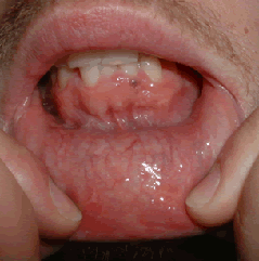
Clinical examination reveals multiple well-demarcated pinpoint-sized dark brown macules of a graded shade of brown (Fig 1). The largest is about 1 mm in diameter and is present in the area apical to teeth #s 23 and 24. There was no induration or palpation sensitivity. The patient had a full set of teeth and fair oral hygiene. Radiographically, tooth #1 was partially impacted, #2 was fully erupted with decay, and #32 was horizontally impacted. Localized periodontal disease in areas adjacent to teeth #s 2 and 31 were identified. There was no evidence of swelling or lymphadenopathy.
Figure 1. This is clinical photograph representing the first clinical presentation where multiple small and pigmented lesions are noted on the anterior mandibular gingiva. The pigmented lesions range in color from tan to very dark brown.
The incisional and excisional surgical specimens were evaluated by Drs. Deubner, Argenyi and Oda. The first two specimens revealed large and atypical melanocytes present along the basal cell layer (Fig 3), some were arranged in small clusters (Fig 4). There was evidence of abnormal mitotic figures and linear spreading of the neoplastic melanocytes (Fig 3), as well as evidence of focal superficial invasion in form of a small cluster of the neoplastic cells. The cells were actively producing and releasing melanin pigment. They were negative with S-100 protein antibody and strongly positive with Melan A antibody (Fig 5). The latter is specific for malignant melanoma. Although unusual for malignant melanoma to be negative with S-100, it has been described in 5% of these lesions.
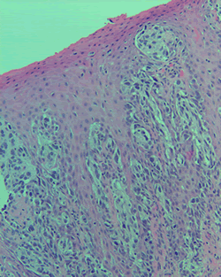
Figure 3. Low Power (x100) histology shows an H & E stained section showing a piece of gingiva with many atypical melanocytes along the rete pegs spreading upwardly and in a linear fashion.
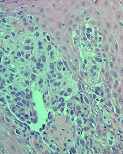
Figure 4. Higher power (x200) histology shows an H & E stained section showing the gingiva with clusters of malignant melanocytes.
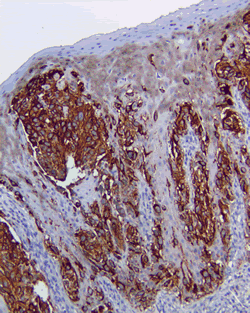
Figure 5. Low power (x100) histology shows the gingiva from Figures 3 and 4 but stained with immunohistochemistry marker Melan A, a specific antibody that stains malignant melanoma cells. Notice the strong positive staining denoted by the dark brown color.
After you have finished reviewing the available diagnostic information