Well-demarcated radiolucency, Anterior Mandible
Can you make the correct diagnosis?
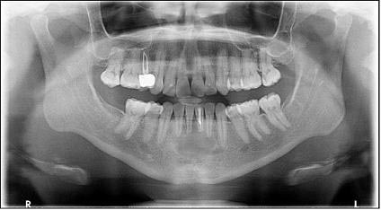
This is a 26-year-old female with some swelling and radiolucency of the anterior mandible of approximately four years’ duration.
Sorry! you are incorrect
Periapical cyst (radicular cyst) or Odontogenic keratocyst (OKC)
Given that the lesion is a large, well-demarcated and corticated unilocular radiolucency, one should think of a cyst first. Since this lesion is large in size and is in the vicinity of, or may be associated with, the apex of an endodontically treated tooth #24, one would think that a periapical cyst is the most likely pathology. The swelling is not unusual for a periapical cyst, especially infected cysts. Another odontogenic cyst to consider is odonotgenic keratocyst (OKC). However, OKC is not usually expansile and would be highly unlikely in this case. It would be unlikely for either of these two types of cysts to cause tooth mobility. The histology is not that of a cyst.
Radicular cyst (periapical cyst, apical periodontal cyst)
Periapical cyst, or radicular cyst, is the most common type of odontogenic cyst. It is inflammatory in origin and is associated with non-vital teeth. Usually, it is apical to the root of a tooth and associated with badly carious teeth, fractured teeth or teeth with large restorations and secondary caries. Periapical cysts can also be lateral to a tooth and can be associated with accessory canals. The maxilla, especially the anterior maxilla, is the most common location. This condition usually occurs in adults over 20 years of age and is typically associated with permanent teeth, but it can be associated with deciduous teeth as well.
Periapical cysts are usually asymptomatic unless infected. They can cause swelling and thinning of the overlying bone to break through the bone in highly infected cases. Radiographically, it is usually unilocular and radiolucent with a corticated border but can be multilocular and have small flecks of calcified material in rare cases. The cortical border can be lost when the cyst is infected. Treatment ranges from extraction of the involved tooth to endodontic filling of the tooth.
Odontogenic keratocyst (OKC)
Odontogenic keratocyst (OKC) is an aggressive odontogenic cyst known for its rapid growth and its tendency to invade the adjacent tissues, including bone. It has a high recurrence rate and is associated with basal cell nevus syndrome. The majority of patients are in the age ranges of 20-29 and 40-59, but cases in patients ranging in age from 5 to 80 years have been reported (4). The distribution between sexes varies from equal distribution to a male-to-female ratio of 1.6:1. OKC predominantly affects Caucasian populations and, if one may judge from the limited evidence provided by the literature, is chiefly of Northern European descent (4). Odontogenic keratocysts may occur in any part of the upper and lower jaw, with the majority (almost 70%) occurring in the mandible. They occur most commonly in the angle of the mandible extending superiorly into the ramus. Radiographically, OKCs present predominantly as unilocular radiolucencies with well-defined or sclerotic borders; they may also present as multilocular radiolucencies or, more commonly, unilocular with scalloped borders. They usually penetrate the bone rather than expand; they grow in an anterior and posterior manner with little to no expansion. The larger OKCs, however, tend to expand bone, but mildly. Obvious clinical expansion should be viewed with suspicion for a neoplasm. OKCs can also present as small and oval radiolucencies between teeth simulating a lateral periodontal cyst, in an area of an extracted tooth simulating a residual cyst, at the apex of a vital tooth mistaken for a periapical cyst, or in the anterior maxilla between the central incisors simulating an incisive canal cyst. OKCs grow to sizes larger than any other odontogenic cysts. Despite this aggressive growth, they often remain asymptomatic, thus growing to large sizes and hollowing the bone. Like other odontogenic cysts, if infected, they can be painful, thus symptomatic. Multiple OKCs are frequently associated with bifid-rib basal cell nevus syndrome (Gorlin syndrome). Odontogenic keratocysts are significant clinical entities due to their tendency for recurrence and destructive behavior. They are known to have a high recurrence rate, ranging from 13% to 60%. Complete surgical removal is the treatment of choice. Enucleation combined with Carnoy’s solution or liquid nitrogen treatment has been effective in reducing recurrence rate.
Sorry! you are incorrect
Again, a well-demarcated unilocular radiolucency should make one think a cyst or a cyst-like structure. The location in the anterior mandible should raise the question of traumatic bone cavity (TBC) as a potential diagnosis. However, the age of the patient is on the older side for TBC, the patient´s gender makes it less likely, and the expansion, although rarely reported, is highly unusual for TBC. The histology is not supportive of this diagnosis either.
The traumatic bone cyst is best called a traumatic bone cavity since this condition does not represent a true cyst. Traumatic bone cavity (TBC) is not unique to the jawbones; it is also described in the long bones and is known as a simple solitary bone cyst occurring mostly in the humerus or femur, close to the epiphyseal plate (6). The long bone simple cyst is similar to the jaw traumatic bone cavity radiographically and occurs in the same age range. Trauma has been suggested as the etiology along with other non-substantiated theories such as cystic degeneration of a preexisting tumor or of the fatty marrow in the area.
Some reports suggest that it is more common in males while others report equal distribution between males and females. The long bone counterpart is more common in males by a ratio of 2.5:1. Most reports agree that the average age of occurrence is below 20 years of age. These lesions can occur, but are uncommon, over the age of 30. Kaugars reported a higher number of TBC cases in African-American females compared to the literature. The latter patients were over the age of 30. This may suggest an association with florid cemento-osseous dysplasia. The mandible is the most commonly affected area, where over 95% of cases occur, especially in the posterior premolar-molar area. They rarely extend to the ramus; therefore, this case is unique in that it extends very high into the ramus. TBCs are also known to cross the midline anteriorly. In one study, 27% of cases were anterior to the canine and some crossed the midline. They are usually unilocular and radiolucent, typically above the alveolar canal, and in many cases have a scalloped superior border spreading between the roots of teeth. The latter are vital and are frequently found hanging within the empty cavity. About 25% of the lesions occur in the anterior mandible apical to the canine tooth and are usually round and unilocular; they can therefore be mistaken for periapical lesions, leading to an unnecessary endondontic treatment. Therefore, it is important to test the vitality of the teeth and carefully examine the radiographs for changes consistent with a periapical granuloma or cyst. Though expansion is not characteristic of TBC, it is described in about 26% of cases. TBCs are otherwise asymptomatic. The margins of these lesions range from very well defined to corticated to punched-out radiolucency. Pathologic fractures associated with traumatic bone cavity have been described in the jaws, but are rare. They are, however, more common in association with TBCs of the long bones.
Clinically, surgeons report an empty cavity at entrance in about two thirds of cases and cavities filled with straw-colored fluid in about one third of cases. Blood clots are also present occasionally. The bony cavity is scraped to generate bleeding, which is considered the treatment of choice for this condition. Other methods of treatment have been tried, such as packing the curetted cavity with autogenous blood, autogenous bone and hydroxyapetite. Various other reports demonstrate healing of TBC after injection of autogenous blood, after aspiration and after endodontic treatment. These lesions may spontaneously heal, but rarely. Biopsy material consists of fragments of viable bone and loose connective tissue. Osteoclast-like giant cells have also been described in a few cases. Exploration surgery usually leads to healing. Recurrence is rare.
Congratulations! You are correct
The age of this patient is not typical for juvenile ossifying fibroma; nor is the location of occurrence. The histology, however, is typical of this condition.
Juvenile ossifying fibroma (JOF) is a rare benign but aggressively behaving neoplasm of fibro-osseous origin that affects the jaws and craniofacial bones. It is an aggressive variant of a more common benign jaw bone neoplasm known as central ossifying fibroma (COF) occurring more commonly in the posterior mandible, inferior to the premolar and molar teeth with a distinct predilection to females at a 5:1 ratio around 35 years of age. Central ossifying fibroma grows in a centrifugal manner in that it can expand the bone bucally and lingually as well as superior and inferior aspects. It is radiographically diagnostic in that it is well demarcated with a corticated border and the central portion of the neoplasm ranges from radiolucent (early stages) to mature radiopacity (late stages) with a connective tissue capsule radiographically identified as a radiolucent rim.
Juvenile ossifying fibroma of the jaws, although a variant of COF, is distinct in several respects: its behavior, the location, the age of the patient and histologic features. As mentioned previously, this neoplasm is locally aggressive. It can simulate the behavior of a low-grade sarcoma with its tendency for fast growth, invading the surrounding tissue and destroying bone, displacing teeth and, at times, causing exophthalmos and diplopia. Tenderness and mild pain is also described, but not parasthesia, as was the case in this patient. It is more common in the maxilla and the craniofacial bones and sinuses.
Two histologic variants of JOF are described: trabecular and psamammatoid types. Trabecular JOF is histologically characterized by cellular connective tissue stroma interspersed with strands of trabeculae young bone with prominent osteoblastic rimming. Psammomatoid JOF is characterized by cellular connective tissue stroma with small and uniform cementum-like hard tissue resembling Psammoma bodies. Trabecular JOF occurs more commonly in the jaw bones; it is about twice as common in the maxilla as in the mandible. The psammamatoid type occurs more commonly in the paranasal sinuses. Both types are suggested to be of equal gender predilection or slightly more common in males. Psamammatoid JOF occurs more commonly in the paranasal sinuses (about 70% of cases) with 20% of cases occurring in the maxilla and 10% in the mandible. Generally, JOF is more common in children under 15 years of age; trabecular JOF tends to occur in younger patients with a range of 8.5-12 years while psamammatoid JOF affects older children with a mean age of 20-22. This patient´s case was more trabecular in type mixed with areas of psammamatoid morphology.
Radiographically, JOF ranges from a well-demarcated radiolucency, as is the case in this patient, to mixed radiolucent/radiopaque or predominantly radiopaque depending on the stage of the disease and degree of calcification. It can be unilocular or multilocular, well defined or ill defined. Both histological types have a high recurrence rate ranging from 30 to 56%. Treatment ranges from thorough curettage to resection to curettage with subcutaneous interferon injections. The latter has been reported to be effective in inhibiting tumor growth and recurrence.
References
- Açikgõz A,Uzun-Bulut E, Ozden B, Gündüz K. Prevalence and distribution of odontogenic and nonodontogenic cysts in a Turkish Population. Med Oral Patol Oral Cir Bucal. 2011 Jul 15.
- Ramachandra P, Maligi P, Raghuveer H. A cumulative analysis of odontogenic cysts from major dental institutions of Bangalore city: A study of 252 cases. J Oral Maxillofac Pathol. 2011 Jan;15(1):1-5.
- Oda D, Rivera V et al. Odontogenic keratocyst: the northwestern USA experience. J Contemp Dent Pract. 2000 Feb 15; 1(2): 60-74.
- Kumar ND, Sherubin JE, Raman U, Shettar S. Solitary bone cyst. Indian J Dent Res. 2011 Jan-Feb;22(1):172-4.
- Kahler B. Traumatic bone cyst suggestive of a chronic periapical abscess: A case report. Aust Endod J. 2011 Aug;37(2):73-5.
- Williams HK, Mangham C, Speight PM. Juvenile ossifying fibroma. An analysis of eight cases and a comparison with other fibro-osseous lesions. J Oral Pathol Med. 2000;29:1318.
- Eversole LR, Leider AS, Nelson K. Ossifying fibroma. A clinicopathologic study of sixty-four cases. Oral Surg Oral Med Oral Pathol. 1985;60:505–511.
- Samir El Mofty Psammomatoid and trabacular juvenile Ossifying Fibroma of the craniofacial skeleton: Two distinct Clinicopathological entities. Oral Surg Oral Med Oral Pathol Oral Radiol Endod 2002; 93:296-304.
- Johnson LC, Yousefi M, Vinh TN, Heffner DK, Hymans VJ and Hartman KS. Juvenile active ossifying fibroma, its Nature, Dynamics and Origin Acta Otolaryngol 1991; suppl 1 488: 1-40.
- Whitaker SB, Vigneswaran N, Budnick SD, Waldron CA. Giant cell lesions of the jaws: evaluation of nucleolar organizer regions of varying behavior. J Oral Pathol Med 1993; 22(9):402-5.
- Tallan EM, Olsen KD, McCaffrey TV, Unni KK, Lund BA. Advanced giant cell granuloma: a twenty-year study. Otolaryngol Head Neck Surg 1994; 110(4):413-8.
- Carlos R, Sedano HO. Intralesional corticosteroids as an alternative treatment for central giant cell granuloma. Oral Surg Oral Med Oral Pathol Oral Radiol Endod 2002; 93(2):161-6.
- O´Regan EM, Gibb DH, Odell EW. Rapid growth of giant cell granuloma in pregnancy treated with calcitonin. Oral Surg Oral Med Oral Pathol Oral Radiol Endod 2001; 92(5):532-8.
- Collins A. Experience with anti-angiogenic therapy of giant cell granuloma of the facial bones. Ann R Australas Coll Dent Surg 2000; 15:170-5.
Sorry! you are incorrect
The age of this patient, the swelling, and the location in the anterior mandible crossing the midline are all clinical characteristics that can be consistent with central giant cell granuloma (CGCG). The histology, however, is not supportive of CGCG.
Jaffe first coined the term “reparative” for central giant cell granuloma. Most pathologists have since dropped the term “reparative” for lack of evidence that the pathogenesis is a reparative process. CGCG is described as a non-neoplastic process and yet can behave in a very aggressive and expansile manner, destroying bone and displacing teeth. Over 60% of CGCG cases occur in patients younger than 30 years of age, with twice as many occurrences in females as in males. CGCG is classified into aggressive and non-aggressive types; the aggressive type tends to occur in younger patients and causes disfiguration, especially after surgery. Over 70% of cases occur in the mandible anterior to the first molar tooth. This lesion has also been described in other cranio-facial and small long bones such as those of the hands and feet.
The usual treatment for CGCG is surgery, ranging from curettage and en bloc to resection. The latter is used in aggressive and recurring cases. In the past ten years or so, alternatives to surgery have emerged with successful results, saving some patients from facial disfigurement. Steroid injections are the most successful alternative treatment thus far; they require injections weekly or every 2-3 weeks, have no known side effects (even in children), and are the least expensive alternative treatment. Other treatments include: calcitonin injections or nasal spray, which require daily injections or a nasal spray of salmon calcitonin for about a year and are safe for pregnant females; and interferon alfa-2a injections, which are administered 2-3 times per week for several months and are the most expensive alternative treatment. A report by Carlos and Sedano from Guatemala presented four patients with large CGCG cases treated with steroid injections. They demonstrated that regular steroid injections led to remarkable responses in all four patients without any side effects. All four patients had initial biopsies confirming the histological diagnosis of CGCG and had endocrine testing to rule out hyperparathyroidism. Two were pediatric patients, two-and-a-half and six years of age, with large lesions of the mandible and maxilla, respectively. They responded more rapidly to steroid injections than did the other two patients, who were adults of 31 and 34 years of age with large lesions of the maxilla and mandible, respectively. Injections were administered every two to three weeks. Complete healing, with small residual radiolucency requiring no further treatment, was noted in three of the patients. One patient, the six-year-old boy, had a maxillary swelling that was reduced from 5 cm in size to only 0.5 cm after four injections. The surgical specimen from the six-year-old consisted of fibrocollagenous stroma with few giant cells in comparison to the original biopsy, which consisted of loose and vascular granulation tissue with many giant cells. Fibrocollagenous stroma, with or without giant cells, has been described in association with both calcitonin and steroid treatment. There was no evidence of recurrence in these patients after 2-7 years of follow-up, nor were there any steroid-related complications in any of the patients. One must keep in mind that this report involves a very small number of cases; few firm conclusions can be reached, but the results are remarkable and show a more favorable outcome than surgical intervention, particularly in large and aggressively behaving pediatric cases.